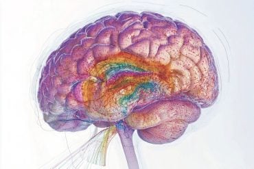
Deadly brain tumors called high-grade gliomas grow with the help of nerve activity in the cerebral cortex, according to a new study by researchers at the Stanford University School of Medicine.
The study, conducted in mice with an aggressive human brain cancer implanted in their brains, is the first to demonstrate stimulation of tumor growth by brain activity. The findings will be published online April 23 in Cell.
It is rare for an organ’s primary function to drive the growth of tumors within it, said Michelle Monje, MD, PhD, senior author of the new study and assistant professor of neurology at the School of Medicine. “We don’t think about bile production promoting liver cancer growth, or breathing promoting the growth of lung cancer,” she said. “But we’ve shown that brain function is driving these brain cancers.”
High-grade gliomas are the leading cause of brain-tumor death in children and adults. Survival rates have scarcely improved in the last 30 years.
“Clinically, fighting high-grade gliomas is a lot like trying to fight a forest fire,” said Monje, who is also a pediatric neuro-oncologist at Lucile Packard Children’s Hospital Stanford, where she cares for patients with these tumors. “Our new findings indicate that this metaphorical forest fire has been difficult to extinguish because there is something akin to gasoline seeping up from the soil.”
Zeroing in on a protein
The tumors studied fell into the broad category of high-grade gliomas: diffuse intrinsic pontine glioma, which strikes school-aged children; pediatric cortical glioblastoma, which affects primarily teens and young adults; anaplastic oligodendroglioma, which affects young adults; and glioblastoma multiforme, which affects older adults. Although these tumors originate in different regions of the brain, all of them originate near or can spread to the cerebral cortex, the brain’s highly folded outer layer that helps us perceive the world, form conscious thoughts and use language.
Monje’s team identified a specific protein, called neuroligin-3, which is largely responsible for the increase in tumor growth associated with neuronal activity in the cerebral cortex. Neuroligin-3 had similar effects across the different types of high-grade gliomas, in spite of the fact that the four cancers have different molecular and genetic characteristics.

“To see a microenvironmental factor that affects all of these very distinct classes of high-grade gliomas was a big surprise,” Monje said.
The identity of the factor was also unexpected. In healthy tissue, neuroligin-3 helps to direct the formation and activity of synapses, playing an important role in the brain’s ability to remodel itself. The new study showed that a secreted form of neuroligin-3 promotes tumor growth.
“This group of tumors hijacks a basic mechanism of neuroplasticity,” Monje said.
Using light stimulation
To conduct the study, Monje’s team employed optogenetics, a Stanford-developed technique that uses genetic manipulation to insert light-sensitive proteins into specific neurons, allowing the neurons to be activated with the flip of a light switch. Into the cerebral cortex of mice with these light-sensitive proteins, the team implanted cancer cells from a human pediatric cortical glioblastoma. After the tumors became established, neurons near the tumors were activated with light. The team then compared tumor growth between these mice and a control group with implanted tumors but without the nerve activation. Increased tumor proliferation and growth in the mice that received neurostimulation via optogenetics were the first indications that neuronal activity fed the brain tumors.
The team performed follow-up experiments on slices of mouse brain to identify secreted factors that made the tumor cells proliferate. They then conducted biochemical analyses to identify neuroligin-3, confirm that the protein could stimulate tumor growth in cultured samples of several kinds of human high-grade gliomas and study which signals the protein uses within glioma cells to promote their growth.
In addition, the researchers examined neuroligin-3 data from The Cancer Genome Atlas, a large public database of human cancer genetics. More activity of the neuroligin-3 gene in high-grade gliomas was linked to shorter survival among patients with these tumors.
The study’s findings may open doors to new high-grade glioma treatments. Although, in theory, the results indicate that sedating patients to reduce neural activity could reduce brain tumor growth, this is unlikely to be accepted as an ethical or practical cancer therapy. A better approach, Monje said, would be to develop drugs that specifically block the tumor-stimulating activities of neuroligin-3, such as a drug that stops the protein from being secreted into the area around the cancer cells.
The lead authors of the study are graduate student Humsa Venkatesh, MD/PhD student Tessa Johung and postdoctoral scholar Viola Caretti, MD, PhD.
Other Stanford co-authors are postdoctoral scholars Erin Gibson, PhD, Yujie Tang, PhD and Jai Pollepalli, PhD; undergraduate student Alyssa Noll; MD/PhD students Surya Nagaraja and Christopher Mount; Siddhartha Mitra, PhD, a senior scientist at the Stanford Institute for Stem Cell Biology and Regenerative Medicine; Pamelyn Woo, life science research assistant; Robert Malenka, MD, PhD, professor of psychiatry and behavioral sciences; Hannes Vogel, MD, professor of pathology and of pediatrics; and Parag Mallick, PhD, assistant professor of radiology.
Monje is also a member of Stanford’s Child Health Research Institute.
Funding: The study was funded by grants from the National Institute of Neurological Disorders and Stroke (grant K08NS070926); the McKenna Claire Foundation; the Matthew Larson Foundation; the National Science Foundation; the Godfrey Family Fund in Memory of Fiona Penelope; the California Institute for Regenerative Medicine; Alex’s Lemonade Stand Foundation; The Cure Starts Now; the Lyla Nsouli Foundation; the Dylan Jewett, Connor Johnson, Zoey Ganesh, Dylan Frick, Abigail Jensen, Wayland Villars, and Jennifer Kranz memorial funds; Unravel Pediatric Cancer; the Virginia and D.K. Ludwig Fund for Cancer Research; the Bear Necessities Pediatric Cancer Foundation; Lucile Packard Foundation for Children’s Health; the Child Health Research Institute at Stanford; and the Anne T. and Robert M. Bass Endowed Faculty Scholarship in Pediatric Cancer and Blood Diseases.
Source: Erin Digitale – Stanford
Image Source: The image is credited to Mikhail Kalinin and is licensed Creative Commons Attribution-ShareAlike 3.0 Unported
Video Source: The video is available at the institutesofmedicine YouTube page
Original Research: Abstract for “Neuronal Activity Promotes Glioma Growth through Neuroligin-3 Secretion” by Humsa S. Venkatesh9 Tessa B. Johung, Viola Caretti, Alyssa Noll, Yujie Tang, Surya Nagaraja, Erin M. Gibson, Christopher W. Mount, Jai Polepalli, Siddhartha S. Mitra, Pamelyn J. Woo, Robert C. Malenka, Hannes Vogel, Markus Bredel, Parag Mallick, and Michelle Monje in Cell. Published online April 23 2015 doi:10.1016/j.cell.2015.04.012
Abstract
Neuronal Activity Promotes Glioma Growth through Neuroligin-3 Secretion
Highlights
•Neuronal activity promotes high-grade glioma (HGG) proliferation and growth
•Neuroligin-3 is an activity-regulated secreted glioma mitogen
•Neuroligin-3 induces PI3K-mTOR signaling in HGG cells
•Neuroligin-3 expression is inversely correlated with survival in human HGG
Summary
Active neurons exert a mitogenic effect on normal neural precursor and oligodendroglial precursor cells, the putative cellular origins of high-grade glioma (HGG). By using optogenetic control of cortical neuronal activity in a patient-derived pediatric glioblastoma xenograft model, we demonstrate that active neurons similarly promote HGG proliferation and growth in vivo. Conditioned medium from optogenetically stimulated cortical slices promoted proliferation of pediatric and adult patient-derived HGG cultures, indicating secretion of activity-regulated mitogen(s). The synaptic protein neuroligin-3 (NLGN3) was identified as the leading candidate mitogen, and soluble NLGN3 was sufficient and necessary to promote robust HGG cell proliferation. NLGN3 induced PI3K-mTOR pathway activity and feedforward expression of NLGN3 in glioma cells. NLGN3 expression levels in human HGG negatively correlated with patient overall survival. These findings indicate the important role of active neurons in the brain tumor microenvironment and identify secreted NLGN3 as an unexpected mechanism promoting neuronal activity-regulated cancer growth.
“Neuronal Activity Promotes Glioma Growth through Neuroligin-3 Secretion” by Humsa S. Venkatesh9 Tessa B. Johung, Viola Caretti, Alyssa Noll, Yujie Tang, Surya Nagaraja, Erin M. Gibson, Christopher W. Mount, Jai Polepalli, Siddhartha S. Mitra, Pamelyn J. Woo, Robert C. Malenka, Hannes Vogel, Markus Bredel, Parag Mallick, and Michelle Monje in Cell. Published online April 23 2015 doi:10.1016/j.cell.2015.04.012


