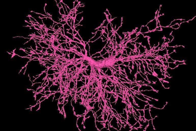
Summary: Oligodendrocyte precursor cells (OPCs) play a significant role in synaptic pruning, a new study reveals.
Source: Allen Institute for Brain Science
JoAnn Buchanan, Ph.D., was deep into the data. One click at a time, she scanned through the branching, twisting 3D shapes of mouse brain cells on her computer screen. Then Buchanan, a scientist at the Allen Institute, saw something weird: a kind of brain cell she wasn’t familiar with.
“They’re just such beautiful cells, they’re so beautiful. And I just became obsessed,” Buchanan said. These brain cells, smaller than neurons but just as complex, are known as oligodendrocyte precursor cells, or OPCs for short.
“I saw something in them which had never been seen before,” Buchanan said.
She doesn’t mean that metaphorically. She literally saw something inside this OPC that nobody else had spotted—machinery that is able to digest parts of other cells.
The OPCs, it turned out, were biting off and swallowing tiny bits of neighboring neurons. It sounds ghoulish, but this is a normal process in the brain. It’s called synaptic pruning and it happens mainly during our brains’ early development.
As neurons form new connections—also called synapses—and lose old ones, other cells in the brain clean up and dispose of the physical machinery from the unwanted connections.
Buchanan and her colleagues published an article describing this new neuron-nibbling role for OPCs in the journal Proceedings of the National Academy of Sciences. Previously, this synapse-junk hauling was thought to be the purview of microglia, a kind of immune cell specialized for the brain.
Microglia are known as the brain’s garbage disposal system—so famous for this task that when Buchanan first discussed her findings with others in the field, some neuroscientists couldn’t believe that OPCs could prune neurons too.
This cellular eating is known as phagocytosis—from the ancient Greek word phagein, which means to consume or devour. Cells that carry out the devouring function are dubbed phagocytes.
“This finding was a complete surprise, because this was believed to be the main purpose of microglia—they’re known as the main phagocytes of the brain,” Buchanan said. “And all of a sudden, here’s this other cell doing it.”
OPCs are known to give rise to another kind of brain cell known as oligodendrocytes, which form thick, white insulation around bundles of nerves. That electrical insulation, also called myelin, makes up the brain’s “white matter” and is also the key brain structure to succumb to damage in the autoimmune disease multiple sclerosis.
But scientists have suspected for a while that OPCs do something else besides generate these insulating brain cells, because there are so many of them in the brain. The Allen Institute team’s findings point to another possible role for these cells.
“These cells are becoming more appreciated now,” said Dwight Bergles, Ph.D., a professor of neuroscience at Johns Hopkins University School of Medicine and a co-author on the OPC study.
“But you would be hard-pressed to find them discussed in neuroscience textbooks, and that’s despite the fact that they make up about 5% of all cells in the central nervous system.”
Something new from the data
Their discovery came about thanks to a massive dataset Buchanan helped create, a collection of the detailed 3D shapes of the nearly 200,000 brain cells present in a grain-of-sand-sized piece of a mouse brain.
That brain cell dataset was generated using an electron microscope, which captures images by bombarding a slice of brain with a beam of focused electrons. Because electrons have a much shorter wavelength than light, electron microscopes can capture tiny details at much higher resolution than light microscopes can.
The kind of electron microscopes used at the Allen Institute pass a beam of electrons through a thin slice of material; the grain of sand sized piece of mouse brain was sliced into 25,000 incredibly thin slices before the team of researchers imaged it.

Buchanan has been working with electron microscopy data her whole career, for more than 40 years now. But she’s never worked with a dataset like this one, she said. For one, there’s the sheer size of it—besides just housing so many cells, it’s large enough to capture complex, branching neurons and other brain cells in their entirety and in relationship with neighboring cells.
But most importantly for the discoveries Buchanan made about OPCs, the dataset has been computationally stitched together from its original super-thin slices into beautiful, 3D visualizations of entire cells.
The project took five years and many teams of scientists to complete. It was carried out in collaboration by the Allen Institute, Baylor College of Medicine, and Princeton University, which generated the computational 3D models that allow for such in-depth exploration.
Now, scientists at all three organizations are gleaning insights from the huge, 3D dataset. Because the data is publicly available, anyone else who wants to can also delve into the thousands of structures inside the tiny speck of mouse brain.
The Allen Institute team has mainly focused on the structures of mouse neurons, which make up slightly less than half of the cells in the dataset, said Nuno Maçarico da Costa, Ph.D., Associate Investigator at the Allen Institute for Brain Science and a co-author on the OPC study. But there’s much more to study in the data than the neurons—like OPCs.
“This finding was only possible with the mixture of JoAnn’s insight and the scale of the dataset,” da Costa said. “She taught us all something new about the data.”
About this neuroscience research news
Author: Press Office
Source: Allen Institute for Brain Science
Contact: Press Office – Allen Institute for Brain Science
Image: The image is credited to Allen Institute
Original Research: Open access.
“Oligodendrocyte precursor cells ingest axons in the mouse neocortex” by JoAnn Buchanan et al. PNAS
Abstract
Oligodendrocyte precursor cells ingest axons in the mouse neocortex
Neurons in the developing brain undergo extensive structural refinement as nascent circuits adopt their mature form. This physical transformation of neurons is facilitated by the engulfment and degradation of axonal branches and synapses by surrounding glial cells, including microglia and astrocytes.
However, the small size of phagocytic organelles and the complex, highly ramified morphology of glia have made it difficult to define the contribution of these and other glial cell types to this crucial process.
Here, we used large-scale, serial section transmission electron microscopy (TEM) with computational volume segmentation to reconstruct the complete 3D morphologies of distinct glial types in the mouse visual cortex, providing unprecedented resolution of their morphology and composition.
Unexpectedly, we discovered that the fine processes of oligodendrocyte precursor cells (OPCs), a population of abundant, highly dynamic glial progenitors, frequently surrounded small branches of axons.
Numerous phagosomes and phagolysosomes (PLs) containing fragments of axons and vesicular structures were present inside their processes, suggesting that OPCs engage in axon pruning. Single-nucleus RNA sequencing from the developing mouse cortex revealed that OPCs express key phagocytic genes at this stage, as well as neuronal transcripts, consistent with active axon engulfment.
Although microglia are thought to be responsible for the majority of synaptic pruning and structural refinement, PLs were ten times more abundant in OPCs than in microglia at this stage, and these structures were markedly less abundant in newly generated oligodendrocytes, suggesting that OPCs contribute substantially to the refinement of neuronal circuits during cortical development.


