

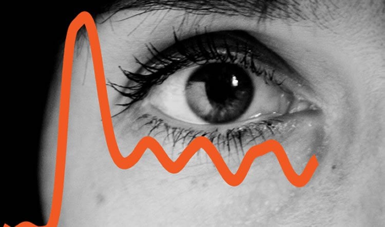
Summary: According to researchers, vision and brain circuits perform regular background scans, making neurons available for focus based tasks. The process makes it possible for us to pay, and maintain, attention.
Source: Newcastle University.
Research reveals that vision and brain circuits perform a regular background scan, making neurons available in case they are needed to focus on a task – enabling us to pay attention.
The work published in Current Biology reveals that this regular visual scan provides a baseline for the brain which later translates into necessary movement, or motor behaviour.
Dr Michael Schmid lead author who is now at Newcastle University, continued this work from the Ernst Strüngmann Institute for Neuroscience in cooperation with the Max Planck Society in Germany. He said: “In simple terms, this scanning function explains for example how we have the mental capacity to react suddenly while driving. This background scanning activity frees up cognitive function, so that if someone steps out in front of the car, we have neurons available to focus on the new action.”
Background scan
The work, carried out in primates, demonstrates that in the tasks studied, the cortex and visual system that make up the attention system, scan the environment every 200 – 300 milliseconds which is 3 – 6 Hz. Other research has detected the same rate in humans.
In-depth analysis of the cortex, revealed that in the network of neurons generating the signals, while one neuron is active, its neighbour would be dormant.
Dr Schmid says: “We believe this shows that we have limited cognitive resources so can’t be paying attention with all our available resources, all the time. This regular background scan ensures that our brains are not overloaded.”
This is a new way to think about attention and could offer new insights into conditions such as Attention deficit hyperactivity disorder (ADHD) and Schizophrenia. The same rhythmic pattern is also observed in reading, so there may be implications for conditions such as dyslexia.
Increasing scientific importance of brain rhythm
Rhythms are an important aspect of brain function. Long believed to be prominent only during sleep, there is increasing evidence that they are also present during wakefulness when engaged in certain tasks. For example, you can experience your rhythmic attention when you engage in a search task such as “Where’s Wally” while observing how your eyes scan the scene. Of course you cannot capture the entire image at the same time, so scanning seems a sensible strategy.
In order to understand the brain rhythm associated with such attentional scanning, the international team recorded the activity of many brain cells in the visual association cortex of monkeys trained to perform a very simple distributed attention task. The monkeys had to execute an eye movement to a target, in this case of a light which would appear unpredictably at one of several possible target locations.
The scientists found the monkey’s reaction times to detect the target followed a theta (3-6 Hz) rhythm which is the same as humans display during such tasks.
In the research, this was shown to be accompanied by a neuronal rhythm before the target was detected. Dr Schmid explains: “We found that the neurons were working together, alternating so that as one neuron became excited, its neighbour was less active.
“We believe that this balance of excitation and inhibition means that there are neurons available if the brain needs to react to a change in circumstance.”
The scientists intend to further the research to understand the mechanism that makes the switch from attentional scanning to focusing, and to reveal the identities of the neurons involved. This may help develop more effective neuropharmacological targeting of attention-supporting drugs.
Source: Newcastle University
Publisher: Organized by NeuroscienceNews.com.
Image Source: NeuroscienceNews.com image is adapted from the Newcastle University news release.
Original Research: Open access research for “Theta Rhythmic Neuronal Activity and Reaction Times Arising from Cortical Receptive Field Interactions during Distributed Attention” by Ricardo Kienitz, Joscha T. Schmiedt, Katharine A. Shapcott, Kleopatra Kouroupaki, Richard C. Saunders, Michael Christoph Schmid in Current Biology. Published July 12 2018.
doi:10.1016/j.cub.2018.05.086
[cbtabs][cbtab title=”MLA”]Newcastle University”Pay Attention: How the Brain Performs a Background Scan to Help Focus.” NeuroscienceNews. NeuroscienceNews, 13 July 2018.
<https://neurosciencenews.com/focus-attention-9569/>.[/cbtab][cbtab title=”APA”]Newcastle University(2018, July 13). Pay Attention: How the Brain Performs a Background Scan to Help Focus. NeuroscienceNews. Retrieved July 13, 2018 from https://neurosciencenews.com/focus-attention-9569/[/cbtab][cbtab title=”Chicago”]Newcastle University”Pay Attention: How the Brain Performs a Background Scan to Help Focus.” https://neurosciencenews.com/focus-attention-9569/ (accessed July 13, 2018).[/cbtab][/cbtabs]
Abstract
Theta Rhythmic Neuronal Activity and Reaction Times Arising from Cortical Receptive Field Interactions during Distributed Attention
Highlights
•Receptive field interactions induce theta rhythmic activation in visual cortex
•The neuronal rhythm does not depend on small fixational eye movements
•Reaction time fluctuations lock to the neuronal rhythm under distributed attention
Summary
Growing evidence suggests that distributed spatial attention may invoke theta (3–9 Hz) rhythmic sampling processes. The neuronal basis of such attentional sampling is, however, not fully understood. Here we show using array recordings in visual cortical area V4 of two awake macaques that presenting separate visual stimuli to the excitatory center and suppressive surround of neuronal receptive fields (RFs) elicits rhythmic multi-unit activity (MUA) at 3–6 Hz. This neuronal rhythm did not depend on small fixational eye movements. In the context of a distributed spatial attention task, during which the monkeys detected a spatially and temporally uncertain target, reaction times (RTs) exhibited similar rhythmic fluctuations. RTs were fast or slow depending on the target occurrence during high or low MUA, resulting in rhythmic MUA-RT cross-correlations at theta frequencies. These findings show that theta rhythmic neuronal activity can arise from competitive RF interactions and that this rhythm may result in rhythmic RTs potentially subserving attentional sampling.


