

Summary: fMRI study reveals dogs do not have a specific face area similar to that of primates. Dogs’ brain activity showed little response to faces but increased in response to seeing another dog over a human.
Source: SfN
Even though dogs gaze into man’s eyes, dog brains may not process faces as human brains do.
A new study from Journal of Neuroscience suggests that the canine visual system is organized differently: the face network found in primates may not extend to all mammals.
Faces constitute a critical part of communication for humans and other primates, so much so that faces have a special status in their visual system. Areas in the face network, like the fusiform face area, activate specifically to faces. Dogs care about faces, too, but they may not have face areas.
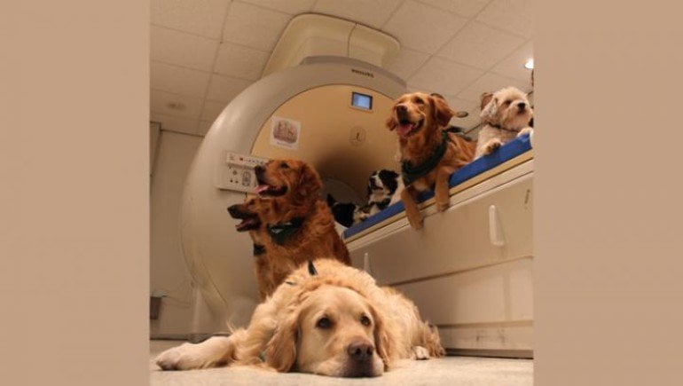
Bunford, Hernández-Pérez et al. used fMRI to compare the brain activity of humans and pet dogs as they watched brief videos of other humans and dogs.
Human brains showed a preference for faces, meaning that some visual areas had greater activity in response to a face compared to the back of the head.
A subset of these regions also displayed species preference, with increased activity in response to viewing a human over a dog.
In contrast, dog brains only showed species preference. Visual areas had greater activity in response to seeing a dog over a human, and no activity difference between seeing a face vs. the back of the head.
About this neuroscience research news
Source: SfN
Contact: Calli McMurray – SfN
Image: Image is credited to Bunford, Hernández-Pérez et al., JNeurosci 2020.
Original Research: Open access.
“Comparative Brain Imaging Reveals Analogous and Divergent Patterns of Species- and Face-Sensitivity in Humans and Dogs” by Nóra Bunford, Raúl Hernández-Pérez, Eszter Borbála Farkas, Laura V. Cuaya, Dóra Szabó, Ádám György Szabó, Márta Gácsi, Ádám Miklósi and Attila Andics. Journal of Neuroscience.
Abstract
Comparative Brain Imaging Reveals Analogous and Divergent Patterns of Species- and Face-Sensitivity in Humans and Dogs
Conspecific-preference in social perception is evident for multiple sensory modalities and in many species. There is also a dedicated neural network for face processing in primates. Yet, the evolutionary origin and the relative role of neural species-sensitivity and face-sensitivity in visuo-social processing are largely unknown. In this comparative study, species- and face-sensitivity to identical visual stimuli (videos of human and dog faces and occiputs) were examined using functional magnetic resonance imaging in dogs (n=20; 45% female) and humans (n=30; 50% female). In dogs, the bilateral mid suprasylvian gyrus showed conspecific-preference, no regions exhibited face-preference, and the majority of the visually-responsive cortex showed greater conspecific- than face-preference. In humans, conspecific-preferring regions (the right amygdala/hippocampus and the posterior superior temporal sulcus) also showed face-preference, and much of the visually-responsive cortex showed greater face- than conspecific-preference. Multivariate pattern analyses identified species-sensitive regions in both species, but face-sensitive regions only in humans. Across-species representational similarity analyses revealed stronger correspondence between dog and human response patterns for distinguishing con- from heterospecific faces than other contrasts. Results unveil functional analogies in dog and human visuo-social processing of conspecificity but suggest that cortical specialization for face perception may not be ubiquitous across mammals.
SIGNIFICANCE STATEMENT
To explore the evolutionary origins of human face-preference and its relationship to conspecific-preference, we conducted the first comparative and noninvasive visual neuroimaging study of a non-primate and a primate species, dogs and humans. Conspecific-preferring brain regions were observed in both species, but face-preferring brain regions were observed only in humans. In dogs, an overwhelming majority of visually-responsive cortex exhibited greater conspecific- than face-preference whereas in humans, much of the visually-responsive cortex showed greater face- than conspecific-preference. Together, these findings unveil functional analogies and differences in the organizing principles of visuo-social processing across two phylogenetically distant mammal species.


