Summary: Results from a new study implicate the ventromedial thalamus in cost-benefit decision making.
Source: OIST
Every day, we make hundreds of decisions. While most are small and inconsequential, like choosing what to eat or wear, others are more complex and involve weighing up potential costs and benefits, like deciding whether to study more for a better grade instead of hanging out with friends.
Decisions like these are shaped by our own values and preferences but understanding how our brains make these choices is still not well understood. Now, scientists at the Okinawa Institute of Science and Technology Graduate University (OIST) have identified a new area of the brain that could be involved in cost-benefit decision-making.
“Previously, many neuroscientists believed that each area of the brain carried out a specific function, such as recognizing faces, memory or movement,” said first author Dr. Bianca Sieveritz, former PhD student and now Junior Research Fellow in the OIST Brain Mechanism for Behavior Unit.
“But in more recent years, we’ve realized that it’s far more complex, and that cognitive processes are carried out by a distributed network across the brain, with many different brain areas communicating.”
To fully understand how cognitive abilities like decision-making work, scientists first need to figure out which parts of the brain are connected to each other. These connections are formed by specialized cells called neurons. Neurons have long, thin projections that can reach out and send signals to other neurons located in different regions of the brain.
But mapping these connections is not easy. “It’s not as simple as identifying that one brain area is connected to another,” explained Dr. Sieveritz.
“Within each brain region, there are many different types, or sub-classes, of neurons, which you need to be able to identify, and each neuronal sub-class might only connect to one other area of the brain or be involved in one specific function. So it’s very messy and complicated.”
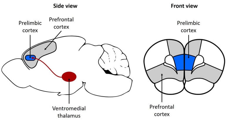
In this study, published in Brain Structure and Function, Dr. Sieveritz examined an area of the brain known as the ventromedial thalamus. This region of the brain is predominantly involved in movement, with abnormal function in this area associated with symptoms of Parkinson’s. But many neuroscientists believe that this area may have other undiscovered functions.
While many neurons from the ventromedial thalamus connect to the motor cortex, other neurons reach further into the prefrontal cortex. This area lies right at the front of our brain and plays a major role in more complex cognitive behaviors, including expression of personality and understanding of language.
But the prefrontal cortex is huge and comprised of millions of neurons, so this research focused in on a tiny area within the prefrontal cortex – the prelimbic cortex. Located right where the two brain hemispheres meet, the prelimbic cortex has previously been found to play a role in fear conditioning, working memory and decision-making.
Working with brain slices from rats, Dr. Sieveritz used different chemical markers to stain the ends of the neurons from the ventromedial thalamus as well as the ends of the different neurons within the outermost layer of the prelimbic cortex. She then pinpointed where the ends of the neurons met each other and formed a connection point, or synapse.
She found that most connections formed within the prelimbic cortex were with a specific sub-class of neurons called cortico-striatal neurons.
“Cortico-striatal neurons in the prelimbic cortex are important for cost-benefit decision-making,” said Dr. Sieveritz.
“If the ventromedial thalamus is sending signals to these cortico-striatal neurons, this could mean that the ventromedial thalamus plays a role in cost-benefit decision-making too.”
Interestingly, Dr. Sieveritz noticed that neurons from the ventromedial thalamus were also sending signals to inhibitory neurons within the prelimbic cortex. Inhibitory neurons slow down or stop other neurons from firing and are essential for keeping the brain’s activity under careful control.

“We know that these inhibitory neurons actually extend down into the cortex and connect to the cortico-striatal neurons in deeper layers of the brain and possibly inhibit their activity,” said Dr. Sieveritz.
“The connections between the neurons from the ventromedial thalamus and the inhibitory neurons therefore may provide a way of finetuning the activity of the cortico-striatal neurons.”
Dr. Sieveritz stressed that while her results implicate the ventromedial thalamus in cost-benefit decision-making, further research is needed to uncover its exact role in this cognitive process.
The team now plans to conduct behavioral studies in rats to determine if signals from the ventromedial thalamus change whether rats choose higher quality food at a higher cost, or lower quality food at a lower cost.
This research brings scientists one step closer to understanding the circuitry underlying the complex process of cost-benefit decision-making.
“Neuroscience is like a huge jigsaw puzzle; everyone can only do a tiny part,” said Dr. Sieveritz. “But when all our research is combined, hopefully we will start to see the bigger picture.”
About this neuroscience research article
Source:
OIST
Contacts:
Tomomi Okubo – OIST
Image Source:
The image is credited to OIST.
Original Research: Open access
“Prelimbic cortical targets of ventromedial thalamic projections include inhibitory interneurons and corticostriatal pyramidal neurons in the rat” by Bianca Sieveritz & Gordon W. Arbuthnott. Brain Structure & Function.
Abstract
Prelimbic cortical targets of ventromedial thalamic projections include inhibitory interneurons and corticostriatal pyramidal neurons in the rat
Ventromedial thalamic axons innervate cortical layer I and make contacts onto the apical dendritic tuft of pyramidal neurons. Optical stimulation of ventromedial thalamic axon terminals in prefrontal cortical areas in mouse brain slices evokes responses in corticocortical, corticothalamic and layer I inhibitory interneurons. Using anterograde tracing techniques and immunohistochemistry in male Sprague–Dawley rats, we provide anatomical evidence that ventromedial thalamic axon terminals in prelimbic cortex make contacts onto pyramidal neurons and, in particular, onto corticostriatal neurons as well as layer I inhibitory interneurons. Using stereology, we made quantitative estimates of contacts in uppermost prelimbic layer I onto dendrites of pyramidal neurons, corticostriatal neurons and layer I inhibitory interneurons. Prefrontal cortex has long been associated with decision making. Specifically, corticostriatal neurons in rat prelimbic cortex play an important role in cost–benefit decision making. Although recent experiments have detailed the physiology of this area in thalamocortical circuits, the extent of the impact of ventromedial thalamic input on corticostriatal neurons or layer I inhibitory interneurons has not been explored. Our quantitative anatomical results provide evidence that most ventromedial thalamic input to pyramidal neurons is provided to corticostriatal neurons and that overall more contacts are made onto the population of excitatory than onto the population of inhibitory neurons.