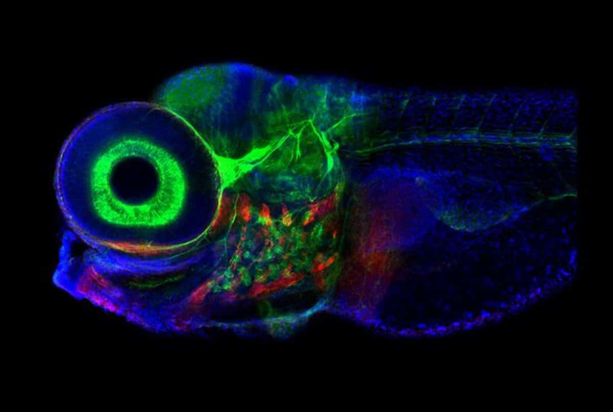
Summary: Researchers used zebrafish larvae to uncover how brainstem neurons guide gaze and maintain short-term memory. By mapping neuronal circuits, they built a computational model that accurately predicted network activity.
The findings shed light on visual-motor systems and offer insights for treating eye movement disorders. Zebrafish, with their simple and accessible neural anatomy, provided a unique opportunity to study brain circuits. This research may pave the way for targeting specific neurons in conditions like eye movement dysfunction.
Key Facts:
- Zebrafish neurons use feedback loops to guide gaze and store short-term memory.
- A computational model of the zebrafish circuit accurately predicted its activity.
- Insights could lead to therapies for eye movement disorders and other brain conditions.
Source: Weill Cornell University
Working with week-old zebrafish larva, researchers at Weill Cornell Medicine and colleagues decoded how the connections formed by a network of neurons in the brainstem guide the fishes’ gaze.
The study, published Nov. 22 in Nature Neuroscience, found that a simplified artificial circuit, based on the architecture of this neuronal system, can predict activity in the network.
In addition to shedding light on how the brain handles short-term memory, the findings could lead to novel approaches for treating eye movement disorders.

Organisms are constantly taking in an array of sensory information about the environment that is changing from one moment to the next.
To accurately assess a situation, the brain must retain these informational nuggets long enough to use them to form a complete picture—for instance, linking together the words in a sentence or allowing an animal to keep its eyes directed to an area of interest.
“Trying to understand how these short-term memory behaviors are generated at the level of neural mechanism is the core goal of the project,” said senior author Dr. Emre Aksay, associate professor of physiology and biophysics at Weill Cornell Medicine, who led the study, together with Dr. Mark Goldman at the University of California Davis and Dr. Sebastian Seung at Princeton University.
Modeling a Dynamical System
To decode the behavior of such neuronal circuits, neuroscientists use the tools of dynamical systems, which involve building mathematical models that describe how the state of a system changes over time, where the current state determines its future states according to a set of rules.
A short-term memory circuit, for example, will remain in a single preferred state until a new stimulus comes along, causing it to settle into a new activity state. In the visual-motor system, each of these states can store the memory of where an animal should be looking.
But what parameters help to set up that type of dynamical system? One possibility is the anatomy of the circuit: the connections that form between each neuron and how many connections they make.
Another likely possibility is physiological strength of those connections, which is established by a myriad of factors like the amount of neurotransmitter being released, the type of synaptic receptors and the concentration of those receptors.
To understand the contributions from circuit anatomy, Dr. Aksay and his collaborators looked to larval zebrafish. By five days of age, these fishlets are swimming around and hunting prey, a skill that involves sustained visual attention.
Importantly for the research team, the brain region that controls eye movement is structurally similar in fish and mammals. But the zebrafish system contains only 500 neurons.
“So, we can analyze the entire circuit—microscopically and functionally,” Dr. Aksay said. “That’s very difficult to do in other vertebrates.”
Zebrafish Shed Light on Neuronal Circuits
Using an array of advanced imaging techniques, Dr. Aksay and colleagues identified the neurons that participate in controlling the animals’ gaze and then determined how these neurons are wired together. They discovered that the system consists of two prominent feedback loops, each containing three clusters of tightly connected cells.
The researchers used this distinctive architecture to build a computational model. They found that their artificial network could accurately predict activity patterns of the zebrafish circuit which they validated by comparing their results to physiological data.
“I consider myself a physiologist, first and foremost,” Dr. Aksay said. “So, I was surprised how much of the behavior of the circuit we could predict from the anatomical architecture alone.”
Next, the researchers will explore how the cells in each cluster contribute to the behavior of the circuit—and whether the neurons in the different clusters have distinct genetic signatures. Such information could allow clinicians to therapeutically target those cells that may malfunction in eye movement disorders.
The findings also provide a blueprint for unraveling the more complex computational systems in the brain that rely on short-term memory, such as those involved in deciphering visual scenes or understanding speech.
Funding: This study was supported in part by the National Institutes of Health grants from the National Institute of Neurological Disorders and Stroke R01 NS104926 and Brain initiative award 5U19NS104648; the National Eye Institute R01 EY027036, R01 EY021581 and K99 EY027017; and the National Cancer Institute UH2 CA203710.
About this neuroscience research news
Author: Corinne Esposito
Source: Weill Cornell University
Contact: Corinne Esposito – Weill Cornell University
Image: The image is credited to Jessica Plavicki
Original Research: Open access.
“Predicting modular functions and neural coding of behavior from a synaptic wiring diagram” by Emre Aksay et al. Nature Neuroscience
Abstract
Predicting modular functions and neural coding of behavior from a synaptic wiring diagram
A long-standing goal in neuroscience is to understand how a circuit’s form influences its function.
Here, we reconstruct and analyze a synaptic wiring diagram of the larval zebrafish brainstem to predict key functional properties and validate them through comparison with physiological data.
We identify modules of strongly connected neurons that turn out to be specialized for different behavioral functions, the control of eye and body movements.
The eye movement module is further organized into two three-block cycles that support the positive feedback long hypothesized to underlie low-dimensional attractor dynamics in oculomotor control.
We construct a neural network model based directly on the reconstructed wiring diagram that makes predictions for the cellular-resolution coding of eye position and neural dynamics.
These predictions are verified statistically with calcium imaging-based neural activity recordings.
This work demonstrates how connectome-based brain modeling can reveal previously unknown anatomical structure in a neural circuit and provide insights linking network form to function.


