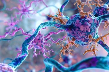
Summary: Researchers report a neural network called the isthmic system helps us to select visual objects that catch our attention.
Source: TUM.
We are almost constantly surrounded by a variety of visual objects, all of which could, theoretically, be important for us. But only a very small area on our retinas, the fovea in the macula lutea, has high visual acuity; a large portion of our field of vision has only a low resolution. Therefore, our gaze must be directed toward a specific target in order to precisely identify the object.
Unconsciously deciding what we look at
But what functionality decides where we direct our vision when we are not looking for anything in particular and do not know where look at in the first place? Researchers from the Department of Zoology at the Weihenstephan School of Life Sciences of the Technical University of Munich, working in cooperation with Chilean and American colleagues, asked themselves this very question.
“The decision where to look is made unconsciously; the objective of our study was to investigate this selection process in detail,” reports Prof. Harald Luksch from the Department of Zoology at the TUM, who also heads the Bionics Center at the TUM. Gaze control involves an evaluation of the field of vision from which a selection is made as to where the fovea will be directed next. “A neural network called the isthmic system performs the selection process,” explains zoologist Luksch. Because this network is well characterized anatomically in birds, the study was carried out on chickens and, in part, on isolated brain tissue (in vitro).
Some stimuli are suppressed, others reinforced
“We were able to demonstrate that individual nerve cells of the visual midbrain establish parallel connections to three areas of the brain,” states Prof. Luksch, summarizing the findings, “which each create their own feedback loops with the visual midbrain.”
This feedback reinforces the most salient visual stimuli while suppressing others at the same time. In this manner, an unconscious selection is made. “What surprised us was that the various feedback loops, reinforcing and inhibitory, are triggered by one and the same cell,” remarks Luksch. “In the past, scientists assumed that this was performed by various cells.” Therefore, a single cell controls entirely different processes, although with different time courses.
Automatic attention control
The importance of this study is not limited to basic research: “The very same mechanisms that work in birds also affect humans in the same way,” says Harald Luksch. This allows us to better understand how our perception and bottom-up attention are controlled, which is, in turn, closely related to our consciousness — one of the most exciting fields of neuroscience.

Because the selection process which has now been researched can be represented as a technical circuit diagram, these intelligently evolved mechanisms in the animal world could also be implemented in robots. It is necessary for robots to react in a manner similar to that of organisms, particularly for interactivity with humans. Therefore, Harald Luksch expects these findings to be important for bionic transfers to technical systems in the future.
Source: Dr. Harald Luksch – TUM
Publisher: Organized by NeuroscienceNews.com.
Image Source: NeuroscienceNews.com image is in the public domain.
Original Research: Abstract for ““Shepherd’s crook” neurons drive and synchronize the enhancing and suppressive mechanisms of the midbrain stimulus selection network” by Florencia Garrido-Charad, Tomas Vega-Zuniga, Cristián Gutiérrez-Ibáñez, Pedro Fernandez, Luciana López-Jury, Cristian González-Cabrera, Harvey J. Karten, Harald Luksch, and Gonzalo J. Marín in PNAS. Published August 7 2018.
doi:10.1073/pnas.1804517115
[cbtabs][cbtab title=”MLA”]TUM”What Catches Our Eye?.” NeuroscienceNews. NeuroscienceNews, 11 September 2018.
<https://neurosciencenews.com/vision-object-selection-9844/>.[/cbtab][cbtab title=”APA”]TUM(2018, September 11). What Catches Our Eye?. NeuroscienceNews. Retrieved September 11, 2018 from https://neurosciencenews.com/vision-object-selection-9844/[/cbtab][cbtab title=”Chicago”]TUM”What Catches Our Eye?.” https://neurosciencenews.com/vision-object-selection-9844/ (accessed September 11, 2018).[/cbtab][/cbtabs]
Abstract
“Shepherd’s crook” neurons drive and synchronize the enhancing and suppressive mechanisms of the midbrain stimulus selection network
The optic tectum (TeO), or superior colliculus, is a multisensory midbrain center that organizes spatially orienting responses to relevant stimuli. To define the stimulus with the highest priority at each moment, a network of reciprocal connections between the TeO and the isthmi promotes competition between concurrent tectal inputs. In the avian midbrain, the neurons mediating enhancement and suppression of tectal inputs are located in separate isthmic nuclei, facilitating the analysis of the neural processes that mediate competition. A specific subset of radial neurons in the intermediate tectal layers relay retinal inputs to the isthmi, but at present it is unclear whether separate neurons innervate individual nuclei or a single neural type sends a common input to several of them. In this study, we used in vitro neural tracing and cell-filling experiments in chickens to show that single neurons innervate, via axon collaterals, the three nuclei that comprise the isthmotectal network. This demonstrates that the input signals representing the strength of the incoming stimuli are simultaneously relayed to the mechanisms promoting both enhancement and suppression of the input signals. By performing in vivo recordings in anesthetized chicks, we also show that this common input generates synchrony between both antagonistic mechanisms, demonstrating that activity enhancement and suppression are closely coordinated. From a computational point of view, these results suggest that these tectal neurons constitute integrative nodes that combine inputs from different sources to drive in parallel several concurrent neural processes, each performing complementary functions within the network through different firing patterns and connectivity.


