Summary: Manipulating blood flow in the brain with visual stimulation induces the flow of cerebrospinal fluid.
Source: PLOS
Researchers at Boston University, USA report that the flow of cerebrospinal fluid in the brain is linked to waking brain activity. Led by Stephanie Williams, and published in the open access journal PLOS Biology on March 30th, the study demonstrates that manipulating blood flow in the brain with visual stimulation induces complementary fluid flow.
The findings could impact treatment for conditions like Alzheimer’s disease, which have been associated with declines in cerebrospinal fluid flow.
Just as our kidneys help remove toxic waste from our bodies, cerebrospinal fluid helps remove toxins from the brain, particularly while we sleep. Reduced flow of cerebrospinal fluid is known to be related to declines in brain health, such as occur in Alzheimer’s disease. Based on evidence from sleep studies, the researchers hypothesized that brain activity while awake could also affect the flow of cerebrospinal fluid.
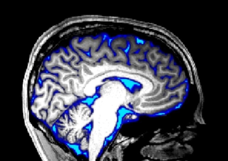
They tested this hypothesis by simultaneously recording human brain activity via fMRI and the speed of cerebrospinal fluid flow while people were shown a checkered pattern that turned on and off.

Researchers first confirmed that the checkered pattern induced brain activity; blood oxygenation recorded by fMRI increased when the pattern was visible and decreased when it was turned off.
Next, they found that the flow of cerebrospinal fluid negatively mirrored the blood signal, increasing when the checkered pattern was off. Further tests showed that changing how long the pattern was visible affected blood and fluid in a predictable way, and that the blood-cerebrospinal fluid link could not be accounted for by only breathing or heart rate rhythms.
Although the study did not measure waste clearance from the brain, it establishes that simple exposure to a flashing pattern can increase the flow of cerebrospinal fluid, which could be a way to combat natural or unnatural declines in fluid flow that occur with age or disease.
Laura Lewis, senior author of the study, adds, “This study discovered that we can induce large changes in cerebrospinal fluid flow in the awake human brain, by showing images with specific patterns. This result identifies a noninvasive way to modulate fluid flow in humans.”
About this neuroscience research news
Author: Press Office
Source: PLOS
Contact: Press Office – PLOS
Image: The image is credited to Stephanie D. Williams (CC-BY 4.0)
Original Research: Open access.
“Neural activity induced by sensory stimulation can drive large-scale cerebrospinal fluid flow during wakefulness in humans” by Laura Lewis et al. PLOS Biology
Abstract
Neural activity induced by sensory stimulation can drive large-scale cerebrospinal fluid flow during wakefulness in humans
Cerebrospinal fluid (CSF) flow maintains healthy brain homeostasis, facilitating solute transport and the exchange of brain waste products. CSF flow is thus important for brain health, but the mechanisms that control its large-scale movement through the ventricles are not well understood.
While it is well established that CSF flow is modulated by respiratory and cardiovascular dynamics, recent work has also demonstrated that neural activity is coupled to large waves of CSF flow in the ventricles during sleep.
To test whether the temporal coupling between neural activity and CSF flow is in part due to a causal relationship, we investigated whether CSF flow could be induced by driving neural activity with intense visual stimulation.
We manipulated neural activity with a flickering checkerboard visual stimulus and found that we could drive macroscopic CSF flow in the human brain. The timing and amplitude of CSF flow was matched to the visually evoked hemodynamic responses, suggesting neural activity can modulate CSF flow via neurovascular coupling.
These results demonstrate that neural activity can contribute to driving CSF flow in the human brain and that the temporal dynamics of neurovascular coupling can explain this effect.


