Each year millions of people worldwide suffer from stroke, which can occur to anyone at any time. While some may recover completely, the majority of survivors will experience some form of impairment that requires a lengthy process toward partial or full recovery of functioning. Therefore, continuous improvement of rehabilitation methods is needed to ensure more positive long-term outcomes among survivors.
Researchers at Japan’s National Institute for Physiological Sciences (NIPS) and Nagoya City University (NCU) have identified the increase of the “cortex-to-red nucleus” pathway via post-stroke focused rehabilitation and its connection to the recovery of functioning in intracerebral hemorrhage (ICH) model rats. This crucial finding could pave the way for better rehabilitative methods for humans.

Stroke often causes hemiplegia by lesioning the corticospinal tract, which connects the motor cortex and spinal cord. Intensive rehabilitation is known to be a promising method of promoting functional recovery via the reorganization of lesioned brain circuits. However, the causal relationship between rehabilitation-induced changes of the brain circuits and functional recovery remains unproven.
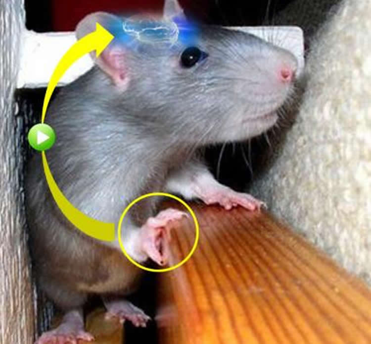
To validate this relationship, the NIPS-NCU team used an ICH rat model to investigate the mechanisms of the therapeutic effects of intensive rehabilitative training. “We found substantial functional recovery of the paralyzed forelimb when applying forced impaired limb use (or FLU) as rehabilitation,” first author Akimasa Ishida explains. “We also uncovered the abundant newly formed connections from the motor cortex to the red nucleus in FLU-treated rats.”
To test the contribution of the formation of new cortex-to-red nucleus connections to functional recovery, the researchers selectively blocked the target pathway using a recently developed double-viral infection technique in rats that experienced recovery. After a blockade of the cortex-to-red nucleus connections, deficiency of the forelimb function reappeared in rehabilitated rats.
The results demonstrate that the rehabilitation-induced reorganization of the damaged brain circuits (i.e., robust increase of the cortex-to-red nucleus connections) is responsible for functional recovery. “We provided direct proof of the causal link between the change of the brain circuits and the recovery of functions by rehabilitation,” corresponding author Tadashi Isa says. “Further investigation on the finer details of the recovery process induced by FLU will help clinicians, physiotherapists, and patients to combat the devastating effects of stroke.”
Source: Tadashi Isa – National Institutes of Natural Sciences
Image Source: The image is credited to Edanz Group Ltd CC-BY-SA 3.0 and is adapted from the National Institutes of Natural Sciences press release.
Original Research: Abstract for “Causal Link between the Cortico-Rubral Pathway and Functional Recovery through Forced Impaired Limb Use in Rats with Stroke” by Akimasa Ishida, Kaoru Isa, Tatsuya Umeda, Kazuto Kobayashi, Kenta Kobayashi, Hideki Hida, and Tadashi Isa in Journal of Neuroscience. Published online January 13 2016 doi:10.1523/JNEUROSCI.2399-15.2016
Abstract
Causal Link between the Cortico-Rubral Pathway and Functional Recovery through Forced Impaired Limb Use in Rats with Stroke
Intensive rehabilitation is believed to induce use-dependent plasticity in the injured nervous system; however, its causal relationship to functional recovery is unclear. Here, we performed systematic analysis of the effects of forced use of an impaired forelimb on the recovery of rats after lesioning the internal capsule with intracerebral hemorrhage (ICH). Forced limb use (FLU) group rats exhibited better recovery of skilled forelimb functions and their cortical motor area with forelimb representation was restored and enlarged on the ipsilesional side. In addition, abundant axonal sprouting from the reemerged forelimb area was found in the ipsilateral red nucleus after FLU. To test the causal relationship between the plasticity in the cortico-rubral pathway and recovery, loss-of-function experiments were conducted using a double-viral vector technique, which induces selective blockade of the target pathway. Blockade of the cortico-rubral tract resulted in deficits of the recovered forelimb function in FLU group rats. These findings suggest that the cortico-rubral pathway is a substrate for recovery induced by intensive rehabilitation after ICH.
SIGNIFICANCE STATEMENT The research aimed at determining the causal linkage between reorganization of the motor pathway induced by intensive rehabilitative training and recovery after stroke. We clarified the expansion of the forelimb representation area of the ipsilesional motor cortex by forced impaired forelimb use (FLU) after lesioning the internal capsule with intracerebral hemorrhaging (ICH) in rats. Anterograde tracing showed robust axonal sprouting from the forelimb area to the red nucleus in response to FLU. Selective blockade of the cortico-rubral pathway by the novel double-viral vector technique clearly revealed that the increased cortico-rubral axonal projections had causal linkage to the recovery of reaching movements induced by FLU. Our data demonstrate that the cortico-rubral pathway is responsible for the effect of intensive limb use.
“Causal Link between the Cortico-Rubral Pathway and Functional Recovery through Forced Impaired Limb Use in Rats with Stroke” by Akimasa Ishida, Kaoru Isa, Tatsuya Umeda, Kazuto Kobayashi, Kenta Kobayashi, Hideki Hida, and Tadashi Isa in Journal of Neuroscience. Published online January 13 2016 doi:10.1523/JNEUROSCI.2399-15.2016


