

Summary: A new study describes the activity of retinal neurons as they deliver visual information to the thalamus, an area of the brain implicated in image processing.
Source: BIDMC.
Most of the human brain’s estimated 86 billion nerve cells, or neurons, can ultimately engage in a two-way dialogue with any other neuron. To shed more light on how neurons in this labyrinthine network integrate information – that is, precisely how multiple neurons send and combine their messages to a target neuron – a team of researchers at BIDMC and Boston Children’s Hospital (BCH) focused on a rare case in which information only travels in one direction: from the retina to the brain.
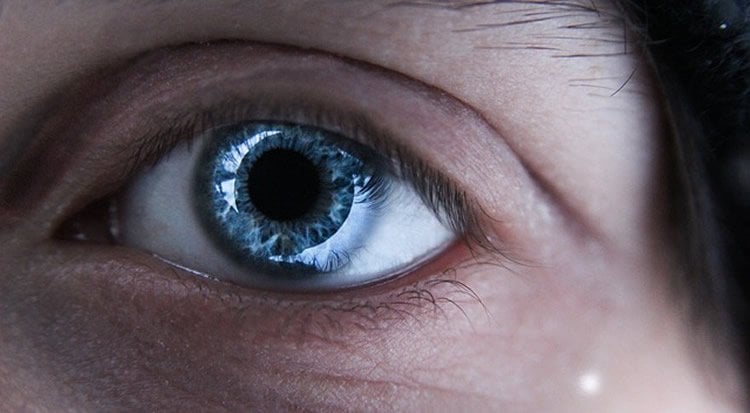
In this study published May 31 in the journal Cell, Mark Andermann, PhD, Chinfei Chen, MD, PhD, and colleagues developed a means of tracking the activity of the far-reaching ends of retinal neurons (called boutons) as they deliver visual information to the thalamus, a brain region involved in image processing.
As they relay discrete bits of visual information to the brain, different types of retinal neurons respond to distinct features of visual content such as an object’s direction of motion, brightness, or size. Conventional wisdom held that these lines of information remained separated in the thalamus. Instead, Andermann and Chen’s team found that boutons from different types of retinal neurons were often organized in local clusters and that boutons in a cluster typically make contact with a common target neuron, leading to a mixing of different lines of information. However, this mixing was not random – boutons in a cluster tended to share a common sensitivity to one or more visual features.
“The selective mixing of information from this arrangement of nearby boutons may be the retina’s version of Pointillism, the neo-expressionist art technique in which nearby dots of different colors are fused together to create new and diverse colors,” said Andermann, a member of the Division of Endocrinology, Diabetes and Metabolism at BIDMC and an Associate Professor of Medicine at Harvard Medical School. “In this way, this first interface between eye and brain is surprisingly sophisticated.”
Other co-investigators included Rohan N. Ramesh, and Arthur U. Sugden, also of BIDMC’s Division of Endocrinology, Diabetes and Metabolism. L. Liang was co-mentored by C. Chen at BCH. RNR, MA and CC also hold appointments in the Program in Neuroscience at Harvard Medical School.
Funding: Support was provided by a Simons Collaboration on the Global Brain Postdoctoral Fellowship (LL), a Bertarelli Foundation Fellowship (AF), NIH F31 105678 (RNR), T32 DK007516 (AUS), R01EY013613 and U54 HD090255 (CC), the Harvard/MIT Joint Research Grants Program in Basic Neuroscience (CC and MA), an NIH Director’s New Innovator Award DP2DK105570, R01 DK109930, and grants from the Smith Family Foundation, the Pew Scholars Program in the Biomedical Sciences (MA), the IDDRC Cellular Imaging Core, and the BCH Viral Core (supported by NIH P30 EY012196).
Source: Lindsey Diaz-MacInnis – BIDMC
Publisher: Organized by NeuroscienceNews.com.
Image Source: NeuroscienceNews.com image is in the public domain.
Original Research: Abstract for “A Fine-Scale Functional Logic to Convergence from Retina to Thalamus” by Liang Liang, Alex Fratzl, Glenn Goldey, Rohan N. Ramesh, Arthur U. Sugden, Josh L. Morgan, Chinfei Chen, and Mark L. Andermann in Cell. Published May 31 2028.
doi:10.1016/j.cell.2018.04.041
[cbtabs][cbtab title=”MLA”]BIDMC”Visualizing the Connections Between Eye and Brain.” NeuroscienceNews. NeuroscienceNews, 2 July 2028.
<https://neurosciencenews.com/eye-brain-connection-9497/>.[/cbtab][cbtab title=”APA”]BIDMC(2028, July 2). Visualizing the Connections Between Eye and Brain. NeuroscienceNews. Retrieved July 2, 2028 from https://neurosciencenews.com/eye-brain-connection-9497/[/cbtab][cbtab title=”Chicago”]BIDMC”Visualizing the Connections Between Eye and Brain.” https://neurosciencenews.com/eye-brain-connection-9497/ (accessed July 2, 2028).[/cbtab][/cbtabs]
Abstract
A Fine-Scale Functional Logic to Convergence from Retina to Thalamus
Highlights
•Chronic imaging reveals diverse visual tuning of mouse retinothalamic boutons
•Clusters of boutons can share one or several visual feature preferences in common
•Functional clustering of boutons occurs at a canonical spatial scale of ∼6 μm
•One axon can innervate multiple clusters specialized for different visual features
Summary
Numerous well-defined classes of retinal ganglion cells innervate the thalamus to guide image-forming vision, yet the rules governing their convergence and divergence remain unknown. Using two-photon calcium imaging in awake mouse thalamus, we observed a functional arrangement of retinal ganglion cell axonal boutons in which coarse-scale retinotopic ordering gives way to fine-scale organization based on shared preferences for other visual features. Specifically, at the ∼6 μm scale, clusters of boutons from different axons often showed similar preferences for either one or multiple features, including axis and direction of motion, spatial frequency, and changes in luminance. Conversely, individual axons could “de-multiplex” information channels by participating in multiple, functionally distinct bouton clusters. Finally, ultrastructural analyses demonstrated that retinal axonal boutons in a local cluster often target the same dendritic domain. These data suggest that functionally specific convergence and divergence of retinal axons may impart diverse, robust, and often novel feature selectivity to visual thalamus.


