

Summary: Using a new technique called amplified MRI, researchers capture the brain in motion as the heart beats.
Source: Stevens Institute of Technology.
Understanding how the brain moves – at rest and upon impact – has been crucial to understanding brain disorders, but technology to clearly see these movements has lagged behind.
Now, researchers from Stevens Institute of Technology, in collaboration with University of Auckland and Stanford University, have developed an imaging technique that captures and magnifies the brain in motion, in real time, every time the heart beats, providing a promising and long-awaited diagnostic tool for catching difficult-to-spot conditions such as concussions and aneurysms – before they become life threatening.
“It’s proof of concept,” says Mehmet Kurt, co-lead author and biomechanical engineer at Stevens. “We wanted to see if we could amplify the tiny movements of the brain with every heartbeat and capture that movement as it naturally occurs – so without introducing noise. That’s important when you are trying to do what we are trying to do: detect abnormal motions in the brain to diagnose and monitor disorders.”
The brain moves slightly with each heartbeat, but these motions are tiny: on the order of ten to 150 micrometers, less than the width of a single human hair. Because these movements are so small, standard MRI techniques have difficulty capturing and displaying them well.
The new technique, to be reported in the May 29 online issue of Magnetic Resonance in Medicine, was originally developed by Samantha Holdsworth and Mahdi Salmani Rahimi while they and Kurt were at Stanford, where they also worked with Itamar Terem. There, Holdsworth and her team developed the foundations for a technique called amplified MRI. In the past two years, the team has fine-tuned the technique, called phase-based amplified MRI, to show that it can be used for diagnostic benefit.
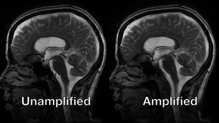
Kurt and Holdsworth attached a pulsometer to healthy subjects and coordinated the timing of the heartbeat with images of the brain and stitched the slices together to create a smooth movement. An algorithm, tailored to the piston-like motions of blood and spinal fluid coursing through the brain, intelligently amplifies the brain’s motion to a more visible scale while keeping potential noise subdued.
The resulting video images, reconstructed slice by slice, retain the spatial characteristics of an MRI – the skull and all anatomical features are displayed at actual scale – while magnifying pulse-driven motion significantly as they animate.
“You can actually capture the whole head ‘nodding’ in the scanner due to the force of the blood pumping into the brain every time the heart beats,” says Holdsworth, a medical physicist who is now at the University of Auckland and co-lead senior author.
Kurt and Holdsworth, along with Terem, found phase-based amplified MRI to produce fewer errors and give better visibility than the original method, particularly areas of the brain that move most, such as the mid-brain and spinal cord, which helps relay sensory information to the brain. The technique also spots movement in areas resistant to motion such as the frontal cortex, which is important for planning, reasoning and judgement.
The team applied the technique on two subjects, a control and a patient with Chiari malformation I. The condition, present at birth, can cause many symptoms, including headaches or stiffness in the neck, due to malformations at the base of the skull and upper spinal area. Unlike the control, video images of the patient showed significantly abnormal brain movement in at least two locations.
Kurt and Holdsworth will continue to advance the technology in clinical settings to larger numbers of patients with known medical diagnoses of various conditions, such as concussion, aneurysm and structural brain abnormalities.
“Better visualization and understanding of the biomechanical properties of the brain could lead to earlier detection and monitoring of brain disorders,” says Kurt, who is also known for his work on concussions. “It could also help with prevention, as it could lead to the design of better helmets.”
Funding: The study was funded by National Institutes of Health, Stanford-Philips Research Agreement, Lucas Foundation.
Source: Thania Benios – Stevens Institute of Technology
Publisher: Organized by NeuroscienceNews.com.
Image Source: NeuroscienceNews.com image is credited to Stevens Institute of Technology.
Original Research: The study will appear in Magnetic Resonance in Medicine.
[cbtabs][cbtab title=”MLA”]Stevens Institute of Technology “Magnifying the Brain in Motion with Every Heartbeat.” NeuroscienceNews. NeuroscienceNews, 29 May 2018.
<https://neurosciencenews.com/brain-motion-heartbeat-9168/>.[/cbtab][cbtab title=”APA”]Stevens Institute of Technology (2018, May 29). Magnifying the Brain in Motion with Every Heartbeat. NeuroscienceNews. Retrieved May 29, 2018 from https://neurosciencenews.com/brain-motion-heartbeat-9168/[/cbtab][cbtab title=”Chicago”]Stevens Institute of Technology “Magnifying the Brain in Motion with Every Heartbeat.” https://neurosciencenews.com/brain-motion-heartbeat-9168/ (accessed May 29, 2018).[/cbtab][/cbtabs]


