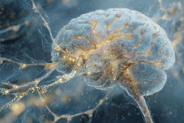
Summary: Researchers use a new imaging technique to confirm structural connections in the brain are unique to each person.
Source: Carnegie Mellon University.
New tool uncovers how brain’s structural connections are individually unique.
Using a new imaging technique, researchers have confirmed what scientists have always thought to be true: the structural connections in the brain are unique to each individual person.
The Carnegie Mellon University-led team used diffusion MRI to map the brain’s structural connections and found each person’s connections are so unique they could identify a person based on this brain “fingerprint” with nearly perfect accuracy. Published in PLOS Computational Biology, the results also show the brain’s that distinctiveness changes over time, which could help researchers determine how factors such as disease, the environment and different experiences impact the brain.
The new, non-invasive diffusion MRI approach captures the brain’s connections at a much closer level than ever before. For example, conventional approaches obtain a single estimate of the integrity of a single structural connection, or a white matter fiber. The new technique measures the integrity along each segment of the brain’s biological wires, making it much more sensitive to unique patterns.
“The most exciting part is that we can apply this new method to existing data and reveal new information that is already sitting there unexplored. The higher specificity allows us to reliably study how genetic and environmental factors shape the human brain over time, thereby opening a gate to understand how the human brain functions or dysfunctions,” said Fang-Cheng (Frank) Yeh, the study’s first author and assistant professor of neurological surgery at the University of Pittsburgh. Yeh completed the research while at CMU as a postdoctoral fellow in psychology.
For the study, the researchers used diffusion MRI to measure the local connectome of 699 brains from five data sets. The local connectome is the point-by-point connections along all of the white matter pathways in the brain, as opposed to the connections between brain regions. To create a fingerprint, they took the data from the diffusion MRI and reconstructed it to calculate the distribution of water diffusion along the cerebral white matter’s fibers.
The measurements revealed that the local connectome is highly unique to an individual and can be used as a personal marker for human identity. To test the uniqueness, the team ran more than 17,000 identification tests. With nearly 100 percent accuracy, they were able to tell whether two local connectomes, or brain “fingerprints,” came from the same person or not.
Additionally, they discovered that identical twins only share about 12 percent of structural connectivity patterns and the brain’s unique local connectome is sculpted over time, changing at an average rate of 13 percent every 100 days.

“This confirms something that we’ve always assumed in neuroscience — that connectivity patterns in your brain are unique to you,” said CMU’s Timothy Verstynen, assistant professor of psychology. “This means that many of your life experiences are somehow reflected in the connectivity of your brain. Thus we can start to look at how shared experiences, for example poverty or people who have the same patholoigical disease, are reflected in your brain connections, opening the door for potential new medical biomarkers for certain health concerns.”
In addition to Yeh and Verstynen, the research team included CMU’s Aarti Singh and Barnabas Poczos, the U.S. Army Research Laboratory’s Jean M. Vettel, the University of California, Santa Barbara’s Scott T. Grafton, the University of Pittsburgh’s Kirk I. Erickson and Wen-Yih I. Tseng of the National Taiwan University.
Funding: The Army Research Laboratory funded this research.
Source: Shilo Rea – Carnegie Mellon University
Image Source: NeuroscienceNews.com image is credited to Carnegie Mellon University.
Original Research: Full open access research for “Quantifying Differences and Similarities in Whole-Brain White Matter Architecture Using Local Connectome Fingerprints” by Fang-Cheng Yeh, Jean M. Vettel, Aarti Singh, Barnabas Poczos, Scott T. Grafton, Kirk I. Erickson, Wen-Yih I. Tseng, Timothy D. Verstynen in PLOS Computational Biology. Published online November 15 2016 doi:10.1371/journal.pcbi.1005203
[cbtabs][cbtab title=”MLA”]Carnegie Mellon University. “A Way to Fingerprint the Brain.” NeuroscienceNews. NeuroscienceNews, 15 November 2016.
<https://neurosciencenews.com/brain-fingerprint-5528/>.[/cbtab][cbtab title=”APA”]Carnegie Mellon University. (2016, November 15). A Way to Fingerprint the Brain. NeuroscienceNews. Retrieved November 15, 2016 from https://neurosciencenews.com/brain-fingerprint-5528/[/cbtab][cbtab title=”Chicago”]Carnegie Mellon University. “A Way to Fingerprint the Brain.” https://neurosciencenews.com/brain-fingerprint-5528/ (accessed November 15, 2016).[/cbtab][/cbtabs]
Abstract
Quantifying Differences and Similarities in Whole-Brain White Matter Architecture Using Local Connectome Fingerprints
Quantifying differences or similarities in connectomes has been a challenge due to the immense complexity of global brain networks. Here we introduce a noninvasive method that uses diffusion MRI to characterize whole-brain white matter architecture as a single local connectome fingerprint that allows for a direct comparison between structural connectomes. In four independently acquired data sets with repeated scans (total N = 213), we show that the local connectome fingerprint is highly specific to an individual, allowing for an accurate self-versus-others classification that achieved 100% accuracy across 17,398 identification tests. The estimated classification error was approximately one thousand times smaller than fingerprints derived from diffusivity-based measures or region-to-region connectivity patterns for repeat scans acquired within 3 months. The local connectome fingerprint also revealed neuroplasticity within an individual reflected as a decreasing trend in self-similarity across time, whereas this change was not observed in the diffusivity measures. Moreover, the local connectome fingerprint can be used as a phenotypic marker, revealing 12.51% similarity between monozygotic twins, 5.14% between dizygotic twins, and 4.51% between none-twin siblings, relative to differences between unrelated subjects. This novel approach opens a new door for probing the influence of pathological, genetic, social, or environmental factors on the unique configuration of the human connectome.
“Quantifying Differences and Similarities in Whole-Brain White Matter Architecture Using Local Connectome Fingerprints” by Fang-Cheng Yeh, Jean M. Vettel, Aarti Singh, Barnabas Poczos, Scott T. Grafton, Kirk I. Erickson, Wen-Yih I. Tseng, Timothy D. Verstynen in PLOS Computational Biology. Published online November 15 2016 doi:10.1371/journal.pcbi.1005203


