Veterans exposed to explosions who do not report symptoms of traumatic brain injury (TBI) may still have damage to the brain’s white matter comparable to veterans with TBI, according to researchers at Duke Medicine and the U.S. Department of Veterans Affairs.
The findings, published in the Journal of Head Trauma Rehabilitation on March 3, 2014, suggest that a lack of clear TBI symptoms following an explosion may not accurately reflect the extent of brain injury.
Veterans of recent military conflicts in Iraq and Afghanistan often have a history of exposure to explosive forces from bombs, grenades and other devices, although relatively little is known about whether this injures the brain. However, evidence is building – particularly among professional athletes – that subconcussive events have an effect on the brain.

“Similar to sports injuries, people near an explosion assume that if they don’t have clear symptoms – losing consciousness, blurred vision, headaches – they haven’t had injury to the brain,” said senior author Rajendra A. Morey, M.D., associate professor of psychiatry and behavioral sciences at Duke University School of Medicine and a psychiatrist at the Durham Veterans Affairs Medical Center. “Our findings are important because they’re showing that even if you don’t have symptoms, there may still be damage.”
Researchers in the Mid-Atlantic Mental Illness Research, Education and Clinical Center at the W.G. (Bill) Hefner Veterans Affairs Medical Center in Salisbury, N.C., evaluated 45 U.S. veterans who volunteered to participate in the study. The veterans, who served since September 2001, were split into three groups: veterans with a history of blast exposure with symptoms of TBI; veterans with a history of blast exposure without symptoms of TBI; and veterans without blast exposure. The study focused on veterans with primary blast exposure, or blast exposure without external injuries, and did not include those with brain injury from direct hits to the head.
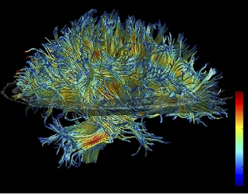
To measure injury to the brain, the researchers used a type of MRI called Diffusion Tensor Imaging (DTI). DTI can detect injury to the brain’s white matter by measuring the flow of fluid in the brain. In healthy white matter, fluid moves in a directional manner, suggesting that the white matter fibers are intact. Injured fibers allow the fluid to diffuse.
White matter is the connective wiring that links different areas of the brain. Since most cognitive processes involve multiple parts of the brain working together, injury to white matter can impair the brain’s communication network and may result in cognitive problems.
Both groups of veterans who were near an explosion, regardless of whether they had TBI symptoms, showed a significant amount of injury compared to the veterans not exposed to a blast. The injury was not isolated to one area of the brain, and each individual had a different pattern of injury.
Using neuropsychological testing to assess cognitive performance, the researchers found a relationship between the amount of white matter injury and changes in reaction time and the ability to switch between mental tasks. However, brain injury was not linked to performance on other cognitive tests, including decision-making and organization.
“We expected the group that reported few symptoms at the time of primary blast exposure to be similar to the group without exposure. It was a surprise to find relatively similar DTI changes in both groups exposed to primary blast,” said Katherine H. Taber, Ph.D., a research health scientist at the W.G. (Bill) Hefner Veterans Affairs Medical Center and the study’s lead author. “We are not sure whether this indicates differences among individuals in symptoms-reporting or subconcussive effects of primary blast. It is clear there is more we need to know about the functional consequences of blast exposures.”
Given the study’s findings, the researchers said clinicians treating veterans should take into consideration a person’s exposure to explosive forces, even among those who did not initially show symptoms of TBI. In the future, they may use brain imaging to support clinical tests.
“Imaging could potentially augment the existing approaches that clinicians use to evaluate brain injury by looking below the surface for TBI pathology,” Morey said.
The researchers noted that the results are preliminary, and should be replicated in a larger study.
Notes about this TBI and neurology research
In addition to Morey and Taber, study authors include Courtney C. Haswell of the Durham VA Medical Center; Susan D. Hurt and Cory D. Lamar of the W.G. (Bill) Hefner VA Medical Center; Jared A. Rowland of the W.G. (Bill) Hefner VA Medical Center and Wake Forest School of Medicine; and Robin A. Hurley of the W.G. (Bill) Hefner VA Medical Center, Wake Forest School of Medicine and Baylor College of Medicine.
This research was supported by a grant from the Department of Defense, Joint Improvised Explosive Device Defeat Organization (51467EGJDO), the Department of Veterans Health Affairs Rehabilitation Research and Development (RX000389-01) and with resources of the Mid-Atlantic Mental Illness Research, Education and Clinical Center and W.G. Hefner VA Medical Center.
Contact: Rachel Harrison – Duke Medicine
Source: Duke Medicine press release
Image Source: The image is credited to Kubicki et al. and is licensed Creative Commons Attribution-Share Alike 3.0 Unported. We adapted the image from the original for this article.
Original Research: Full open access research (PDF) for “White Matter Compromise in Veterans Exposed to Primary Blast Forces” by Katherine H. Taber, PhD; Robin A. Hurley, MD; Courtney C. Haswell, MS; Jared A. Rowland, PhD; Susan D. Hurt, PhD; Cory D. Lamar, MD; and Rajendra A. Morey, MD in Journal of Head Trauma Rehabilitation. Published online March 3 2014 doi:10.1097/HTR.0000000000000030


