Summary: Researchers have discovered specific neurochemical changes in brain regions associated with brain remapping following amputation. Also, these changes may persist once the limb is reattached.
Source: University of Missouri Columbia.
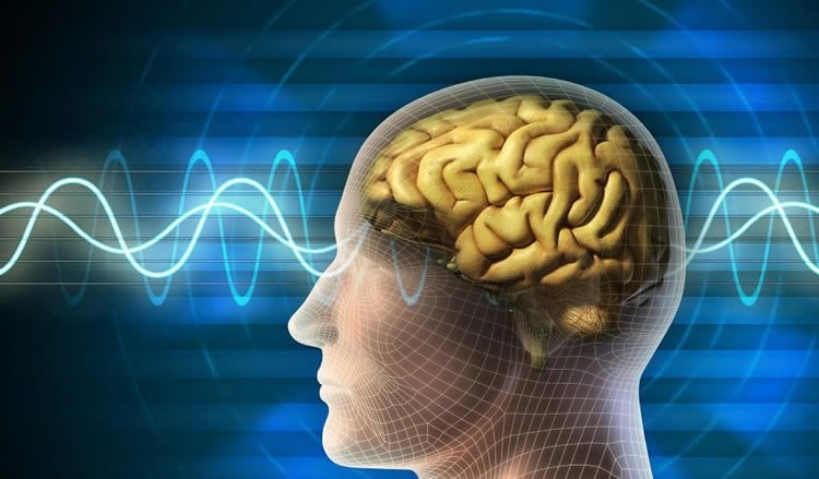
When a person loses a hand to amputation, nerves that control sensation and movement are severed, causing dramatic changes in areas of the brain that controlled these functions. As a result, areas of the brain devoted to the missing hand take on other functions. Now, researchers from the University of Missouri have found evidence of specific neurochemical changes associated with lower neuronal health in these brain regions. Further, they report that some of these changes in the brain may persist in individuals who receive hand transplants, despite their recovered hand function.
“When there is a sudden increase or decrease in stimulation that the brain receives, the function and structure of the brain begins to change,” said Carmen M. Cirstea, M.D., Ph.D., research assistant professor of Physical Medicine and Rehabilitation and lead author of the study. “Using a noninvasive approach known as magnetic resonance spectroscopy (MRS) to examine areas of the brain previously involved with hand function, we observed the types of changes taking place at the neurochemical level after amputation, transplantation or reattachment.”
Cirstea, with co-author Scott Frey, Ph.D., the Miller Family Chair in Cognitive Neuroscience in the Departments of Psychological Sciences and Neurology, used MRS to evaluate the neuronal health and function of nerve cells of current hand amputees, former amputees and healthy subjects.
The researchers instructed volunteers to flex their fingers to activate sensorimotor areas in both sides of the brain. The research team then analyzed N-acetylaspartate (NAA) levels, a chemical associated with neuronal health. The researchers found that NAA values for the reattachment and transplant patients were similar to levels of amputees and significantly lower than the healthy control group.
“Previous research has found substantial reorganizational changes in the brain following limb injuries that decrease sensory and motor stimulation following limb injuries,” Frey said. “These findings show that after surgical repairs, the effects of nerve injuries on the mature brain may continue even as former amputees recover varying degrees of sensory and motor functions in replanted or transplanted hands.”
Due to the small number of reattachment and transplant patients studied (5), the researchers said that the results should be interpreted with caution until more work is completed.
The study, “Magnetic Resonance Spectroscopy of Current Hand Amputees Reveals Evidence for Neuronal-level Changes in Former Sensorimotor Cortex,” was published in the Journal of Neurophysiology, and was recognized by the American Physiological Society as one of the top original research papers published in April 2017.
Funding: Research reported in this publication was supported by the United States Army Medical Research Acquisition Activity Grant, the National Institute of Neurological Disorders and Stroke Grant, the National Institutes of Health and the U.S. Department of Defense. The researchers have no conflicts of interest to declare related to this study.
Source: University of Missouri Columbia
Image Source: NeuroscienceNews.com image is adapted from the UM news release.
Original Research: Full open access research for “Magnetic resonance spectroscopy of current hand amputees reveals evidence for neuronal-level changes in former sensorimotor cortex” by Carmen M. Cirstea, In-Young Choi, Phil Lee, Huiling Peng, Christina L. Kaufman, and Scott H. Frey in Journal of Neurophysiology. Published online April 1 2017 doi:10.1152/jn.00329.2016
[cbtabs][cbtab title=”MLA”]University of Missouri Columbia “How Hand Amputation and Reattachment Affect the Brain.” NeuroscienceNews. NeuroscienceNews, 24 May 2017.
<https://neurosciencenews.com/hand-amputation-reattachment-6762/>.[/cbtab][cbtab title=”APA”]University of Missouri Columbia (2017, May 24). How Hand Amputation and Reattachment Affect the Brain. NeuroscienceNew. Retrieved May 24, 2017 from https://neurosciencenews.com/hand-amputation-reattachment-6762/[/cbtab][cbtab title=”Chicago”]University of Missouri Columbia “How Hand Amputation and Reattachment Affect the Brain.” https://neurosciencenews.com/hand-amputation-reattachment-6762/ (accessed May 24, 2017).[/cbtab][/cbtabs]
Abstract
Magnetic resonance spectroscopy of current hand amputees reveals evidence for neuronal-level changes in former sensorimotor cortex
Deafferentation is accompanied by large-scale functional reorganization of maps in the primary sensory and motor areas of the hemisphere contralateral to injury. Animal models of deafferentation suggest a variety of cellular-level changes including depression of neuronal metabolism and even neuronal death. Whether similar neuronal changes contribute to patterns of reorganization within the contralateral sensorimotor cortex of chronic human amputees is uncertain. We used functional MRI-guided proton magnetic resonance spectroscopy to test the hypothesis that unilateral deafferentation is associated with lower levels of N-acetylaspartate (NAA, a putative marker of neuronal integrity) in the sensorimotor hand territory located contralateral to the missing hand in chronic amputees (n = 19) compared with the analogous hand territory of age- and sex-matched healthy controls (n = 28). We also tested whether former amputees [i.e., recipients of replanted (n = 3) or transplanted (n = 2) hands] exhibit NAA levels that are indistinguishable from controls, possible evidence for reversal of the effects of deafferentation. As predicted, relative to controls, current amputees exhibited lower levels of NAA that were negatively and significantly correlated with the time after amputation. Contrary to our prediction, NAA levels in both replanted and transplanted patients fell within the range of the current amputees. We suggest that lower levels of NAA in current amputees reflects altered neuronal integrity consequent to chronic deafferentation. Thus local changes in NAA levels may provide a means of assessing neuroplastic changes in deafferented cortex. Results from former amputees suggest that these changes may not be readily reversible through reafferentation.
NEW & NOTEWORTHY This study is the first to use functional magnetic resonance-guided magnetic resonance spectroscopy to examine neurochemical mechanisms underlying functional reorganization in the primary somatosensory and motor cortices consequent to upper extremity amputation and its potential reversal through hand replantation or transplantation. We provide evidence for selective alteration of cortical neuronal integrity associated with amputation-related deafferentation that may not be reversible.
“Magnetic resonance spectroscopy of current hand amputees reveals evidence for neuronal-level changes in former sensorimotor cortex” by Carmen M. Cirstea, In-Young Choi, Phil Lee, Huiling Peng, Christina L. Kaufman, and Scott H. Frey in Journal of Neurophysiology. Published online April 1 2017 doi:10.1152/jn.00329.2016