Summary: New research provides some details on the characteristics of motor detection in the brains of flies.
Source: Max Planck Institute.
Neurobiologists discover important characteristics of the motion detector in the fly brain.
In order to react to changes in the environment in good time, the brain must analyze the signals it receives from the eyes rapidly and accurately. For example, the ability to recognise the direction in which an approaching car is moving is vital to the survival of modern humans in cities. Using the brain of the fruit fly Drosophila as a model, scientists from the Max Planck Institute of Neurobiology study how the brain extracts this essential motion information. They have now described in detail the cells that enable downstream neurons to recognize the direction of movement. Interestingly, the characteristics of these input cells exactly match to a motion detector model they recently proposed. In addition, the cells alter their characteristics according to the animals’ state: when the fly is active, the cells respond faster to light stimuli.
Humans perceive their environment mainly through their eyes. The ability to recognize movements and their direction is something that seems almost trivial and automatic to us. However, this information has to be processed in the brain as the light-sensitive sensory cells of the retina can only register changes in contrast. The direction of a movement can only be calculated through the comparison of neighbouring signals. Various models exist for these calculations. Alexander Borst and his team at the Max Planck Institute of Neurobiology study the extent to which these models can be applied to the brain’s neuronal circuitry. Their test subject is the fruit fly Drosophila, a master of motion perception.
In the brain of the fruit fly, the T4 and T5 cells are the first neurons that respond to a motion stimulus in a direction-selective way. It was long assumed that these cells compare the signals from two adjacent image points in the fly’s field of vision and in this way calculate the direction of motion. However, this very simple model was unable to explain all of the empirical measurements recorded by the neurobiologists. “So our department developed a model in which three rather than two adjacent image points are compared with each other,” says Alexander Arenz, explaining the starting point of the current study. “We wanted to test this model using the cells in the fly brain.” A new anatomical study had revealed beforehand that both T4 and T5 cells receive their input signals from four different types of cells. Hence a model with three input cells appeared plausible.
Using a kind of fly cinema, the researchers presented different light stimuli to the flies and recorded the reactions of the different input cell types. The results revealed strong variations in the speed and duration of the individual cells’ responses to changes in brightness. To analyze the significance of these differences in greater detail, the researchers fed the measured data on the cell characteristics into a computer simulation of the network. The simulations indicated that the measured differences between the cells and the resulting delays with which the T4 and T5 cells receive their signals are essential for the recognition of a direction of motion. “So the motion detector with three cells functions very well with these cells,” explains Michael Drews, one of the study’s two first authors. “We now have to find out how the fourth input cell gets involved in this circuit,” he adds.

To narrow down the tasks of the individual cells even further, the researchers availed of the fact that the fly brain processes visual impressions faster when the animal moves. “This can be expected, as the surroundings move past the eyes far more rapidly when in motion,” explains Alexander Arenz. To generate this state in the fly brain, the scientists stimulated octopamine receptors, which are activated under natural conditions when the animals move. The measurements they recorded showed that under these “active conditions” the input cells worked faster and responded more strongly to higher motion speeds – and, as a result, the downstream T4 and T5 cells did so, too.
“The results demonstrate impressively the flexibility with which the neuronal motion detectors can adapt to the animals’ behavioural state”, explains Alexander Borst in summary. In this way the fruit fly can reliably perceive movements in its environment when it moves at a high speed. No wonder flies are so hard to catch.
Source: Stefanie Merker – Max Planck Institute
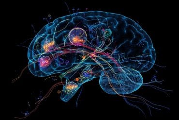
Image Source: NeuroscienceNews.com image is credited to MPI of Neurobiology / Meier, Serbe & Arenz.
Original Research: Abstract for “The temporal tuning of the Drosophila motion detectors is determined by the dynamics of their input elements” by Alexander Arenz, Michael S. Drews, Florian G. Richter, Georg Ammer and Alexander Borst in Nature Chemical Biology. Published online March 23 2017 doi:10.1016/j.cub.2017.01.051
[cbtabs][cbtab title=”MLA”]Max Planck Institute “The More Active the Fly, the Faster Its Brain Works.” NeuroscienceNews. NeuroscienceNews, 3 April 2017.
<https://neurosciencenews.com/fly-activity-brain-6334/>.[/cbtab][cbtab title=”APA”]Max Planck Institute (2017, April 3). The More Active the Fly, the Faster Its Brain Works. NeuroscienceNew. Retrieved April 3, 2017 from https://neurosciencenews.com/fly-activity-brain-6334/[/cbtab][cbtab title=”Chicago”]Max Planck Institute “The More Active the Fly, the Faster Its Brain Works.” https://neurosciencenews.com/fly-activity-brain-6334/ (accessed April 3, 2017).[/cbtab][/cbtabs]
Abstract
The temporal tuning of the Drosophila motion detectors is determined by the dynamics of their input elements
Highlights
•Input neurons to the fly motion detectors show diverse temporal filter properties
•Octopamine activation accelerates detector velocity tuning and input cell dynamics
•Dynamics of input neurons correctly predict velocity tuning of motion detectors
Summary
Detecting the direction of motion contained in the visual scene is crucial for many behaviors. However, because single photoreceptors only signal local luminance changes, motion detection requires a comparison of signals from neighboring photoreceptors across time in downstream neuronal circuits. For signals to coincide on readout neurons that thus become motion and direction selective, different input lines need to be delayed with respect to each other. Classical models of motion detection rely on non-linear interactions between two inputs after different temporal filtering. However, recent studies have suggested the requirement for at least three, not only two, input signals. Here, we comprehensively characterize the spatiotemporal response properties of all columnar input elements to the elementary motion detectors in the fruit fly, T4 and T5 cells, via two-photon calcium imaging. Between these input neurons, we find large differences in temporal dynamics. Based on this, computer simulations show that only a small subset of possible arrangements of these input elements maps onto a recently proposed algorithmic three-input model in a way that generates a highly direction-selective motion detector, suggesting plausible network architectures. Moreover, modulating the motion detection system by octopamine-receptor activation, we find the temporal tuning of T4 and T5 cells to be shifted toward higher frequencies, and this shift can be fully explained by the concomitant speeding of the input elements.
“The temporal tuning of the Drosophila motion detectors is determined by the dynamics of their input elements” by Alexander Arenz, Michael S. Drews, Florian G. Richter, Georg Ammer and Alexander Borst in Nature Chemical Biology. Published online March 23 2017 doi:10.1016/j.cub.2017.01.051


