Summary: Exposure to phobic images without conscious awareness appears to produce more effective results than conscious exposure when it comes to helping people reduce their fear, a new study reports.
Source: Children’s Hospital of Los Angeles.
A team of investigators, led by Bradley S. Peterson, MD, director of the Institute for the Developing Mind at Children’s Hospital Los Angeles, and Paul Siegel, PhD, associate professor of psychology at Purchase College of the State University of New York, have found that exposure to phobic images without conscious awareness is more effective than longer, conscious exposure for reducing fear. The investigators used fMRI to determine that areas of the brain involved in fear processing were much more strongly activated by unconscious exposure. Results of the study will be published in the journal, Human Brain Mapping, on February 6, 2017.
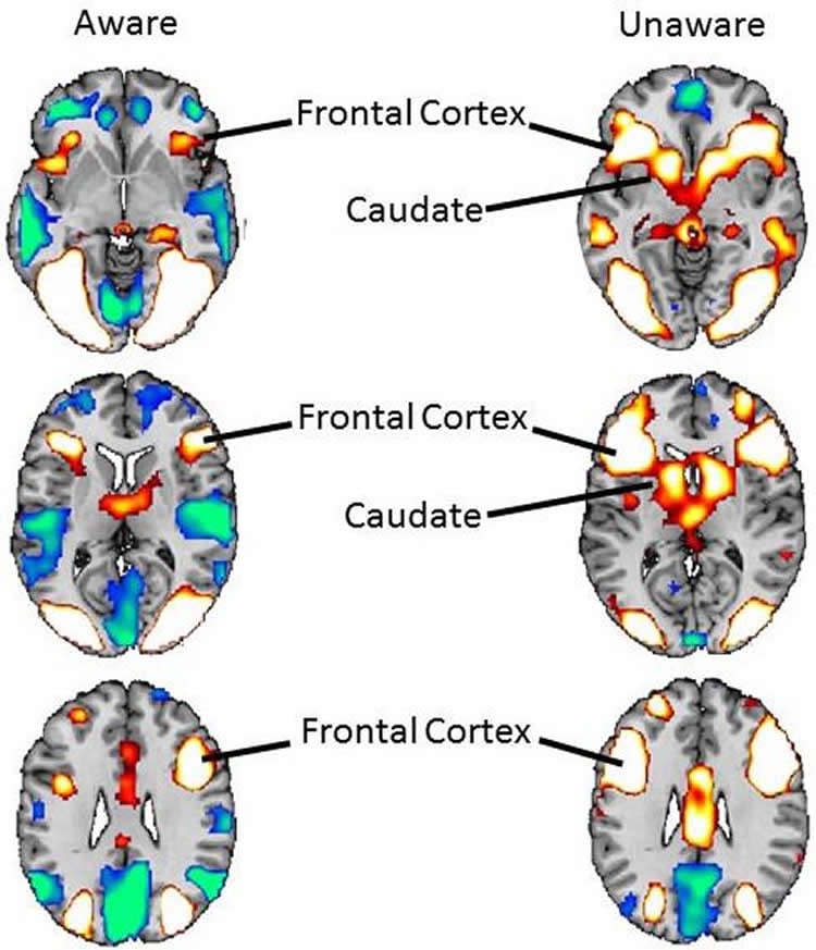
“Although we expected – and observed – activation of the neural regions that process fear,” said Peterson, who is also professor of pediatrics and psychiatry at the Keck School of Medicine of the University of Southern California, “we also found activation in regions that regulate the emotional and behavioral responses to fear–reducing the conscious experience of fear.”
While “phobia” is often defined as an irrational fear, many of the stimuli that produce a phobic response actually have an evolutionary basis that biologically prepares humans to fear them. For this study, the investigators used spiders – a common fear stimulus. They enrolled 21 spider-phobic and 21 non-phobic control participants, all young adult women. Women were selected because previous research has shown that 75 to 80 percent of all people who experience phobias are women.
All participants experienced three conditions that included viewing control images not associated with phobias (flowers) and phobia-inducing images (spiders) at two levels of exposure – very brief (without awareness) and longer duration (clearly visible). The very brief exposure was accomplished through a technique known as backward masking, where a target image is shown very briefly and then immediately followed by a non-target image or “mask” that prevents recognition of the target.
In participants with phobia, very brief exposure to spider images strongly activated the subcortical regions of the brain involved in immediate fear processing. Yet they did not experience fear consciously, apparently because the very brief exposures also activated brain regions that regulate fear. Clearly visible exposure to the spider images, by contrast, deactivated areas of the brain that regulate fear responses, inducing the conscious experience of fear.
“Counter-intuitively, our study showed that the brain is better able to process feared stimuli when they are presented without conscious awareness,” said Siegel, who is first author on the study. “Our findings suggest that phobic people may be better prepared to face their fears if at first they are not consciously aware that they’ve faced them.”
Peterson added that he saw potential for using this technique to treat children and adolescents with anxiety disorders. Current therapies are based on directly confronting the feared stimulus, which can cause young people to experience significant emotional distress.
Additional contributors to the study include Richard Warren and Lilly Murray, Purchase College, SUNY; Zhishun Wang and Jie Yang, Columbia University; Don Cohen, New York University; and Jason Anderson, University of California, Santa Barbara.
Funding: The study was funded by a grant from the National Institute of Mental Health (7R21MH102564-02). Supplemental funding was provided by the American Psychoanalytic Association, the Society for Neuropsychoanalysis, the International Psychoanalytic Association and Purchase College.
Source: Ellin Kavanagh – Children’s Hospital of Los Angeles
Image Source: NeuroscienceNews.com image is credited to Bradley Peterson, MD.
Original Research: Abstract for “Less is more: Neural activity during very brief and clearly visible exposure to phobic stimuli” by Paul Siegel, Richard Warren, Zhishun Wang, Jie Yang, Don Cohen, Jason F. Anderson, Lilly Murray and Bradley S. Peterson in Human Brain Mapping. Published online February 6 2017 doi:10.1002/hbm.23533
[cbtabs][cbtab title=”MLA”]Children’s Hospital of Los Angeles “Emapthetic People Experience Dogs’ Expressions More Strongly.” NeuroscienceNews. NeuroscienceNews, 6 February 2017.
<https://neurosciencenews.com/exposure-stimuli-phobias-6066/>.[/cbtab][cbtab title=”APA”]Children’s Hospital of Los Angeles (2017, February 6). Emapthetic People Experience Dogs’ Expressions More Strongly. NeuroscienceNew. Retrieved February 6, 2017 from https://neurosciencenews.com/exposure-stimuli-phobias-6066/[/cbtab][cbtab title=”Chicago”]Children’s Hospital of Los Angeles “Emapthetic People Experience Dogs’ Expressions More Strongly.” https://neurosciencenews.com/exposure-stimuli-phobias-6066/ (accessed February 6, 2017).[/cbtab][/cbtabs]
Abstract
Less is more: Neural activity during very brief and clearly visible exposure to phobic stimuli
Research on automatic processes in fear has emphasized the provocation of fear responses rather than their attenuation. We have previously shown that the repeated presentation of feared images without conscious awareness via backward masking reduces avoidance of a live tarantula in spider-phobic participants. Herein we investigated the neural basis for these adaptive effects of masked exposure. 21 spider-phobic and 21 control participants, identified by a psychiatric interview, fear questionnaire, and approaching a live tarantula, viewed stimuli in each of three conditions: (1) very brief exposure (VBE) to masked images of spiders, severely limited awareness; (2) clearly visible exposure (CVE) to spiders, full awareness; and (3) masked images of flowers (control), severely limited awareness. Only VBE to masked spiders generated neural activity more strongly in phobic than in control participants, within subcortical fear, attention, higher-order language, and vision systems. Moreover, VBE activated regions that support fear processing in phobic participants without causing them to experience fear consciously. Counter-intuitively, CVE to the same spiders generated stronger neural activity in control rather than phobic participants within these and other systems. CVE deactivated regions supporting fear regulation and caused phobic participants to experience fear. CVE-induced activations also correlated with measures of explicit fear ratings, whereas VBE-induced activations correlated with measures of implicit fear (color-naming interference of spider words). These multiple dissociations between the effects of VBE and CVE to spiders suggest that limiting awareness of exposure to phobic stimuli through visual masking paradoxically facilitates their processing, while simultaneously minimizing the experience of fear.
“Less is more: Neural activity during very brief and clearly visible exposure to phobic stimuli” by Paul Siegel, Richard Warren, Zhishun Wang, Jie Yang, Don Cohen, Jason F. Anderson, Lilly Murray and Bradley S. Peterson in Human Brain Mapping. Published online February 6 2017 doi:10.1002/hbm.23533