

Summary: Using neuroimaging and mathematical graph theory, researchers discover it is possible to detect those at risk of developing psychosis by examining cortical folding.
Source: University of Basel.
Imaging techniques can be used to detect the development of psychosis in the brains of high-risk patients at an early stage, as reported by researchers from the University of Basel and Western University in the journal JAMA Psychiatry.
Detecting psychosis early increases the chances of effective treatment. Despite advances in diagnosis, however, it has previously not been possible to examine young people with initial psychotic symptoms and reliably say who will develop acute psychosis and who will not.
It has long been supposed that the condition is caused by disturbed communication between various groups of nerve cells. Modern imaging techniques have made it possible to make these connections between regions of the brain visible.
Researchers from the University of Basel have now examined the question of whether changes in the anatomical structure of brain networks can already be detected in people with an increased risk of psychosis. The study was carried out in collaboration with scientists from the Psychiatric University Clinics Basel, Western University and Lawson Health Research Institute in Ontario, Canada.
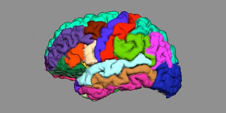
Focus on cortical folding
The researchers, led by Drs André Schmidt and Lena Palaniyappan, focused on cortical folding, known as gyrification: they examined how the folds in various regions of the brain interact with each other, and whether this interaction is impaired in high-risk patients.
They also tested how precisely they could use the cortical connectivity to predict which high-risk patients would suffer from psychosis and which would not.
Reduced interaction
For their study, the researchers examined 44 healthy control subjects, 38 patients with first-episode psychosis, and 79 people with an increased risk of psychosis, of which 16 later developed fully-formed psychosis. They reconstructed the brain’s nerve pathways using magnetic resonance imaging and methods from mathematical graph theory, with which they described a network of nodes.
The results show that in comparison to the healthy control group, the folding in individual regions of the brain in patients with an initial psychotic episode and those with a later psychosis transition showed reduced integration and increased segregation.
The analysis also showed that this process enabled predictions to be made with more than 80% accuracy about which patients would later suffer from psychosis and which would not.
Biomarker for clinical diagnosis
“Our results indicate that this type of network analysis could significantly improve individual risk prognoses,” says André Schmidt, who led the project. “Future longitudinal studies with larger samples are now needed to validate the prognostic accuracy of this measurement.”
Source: Cornelia Niggli – University of Basel
Publisher: Organized by NeuroscienceNews.com.
Image Source: NeuroscienceNews.com image is credited to University Psychiatric Clinics Basel.
Original Research: Open access research for “Disorganized Gyrification Network Properties During the Transition to Psychosis” by Tushar Das, PhD; Stefan Borgwardt, MD; Daniel J. Hauke, MSc; Fabienne Harrisberger, PhD; Undine E. Lang, MD; Anita Riecher-Rössler, MD; Lena Palaniyappan, MD; André Schmidt, PhD in JAMA Psychiatry. Published April 25 2018.
doi:10.1001/jamapsychiatry.2018.0391
[cbtabs][cbtab title=”MLA”]University of Basel “Indications of Psychosis Appear in Cortical Folding.” NeuroscienceNews. NeuroscienceNews, 25 April 2018.
<https://neurosciencenews.com/psychosis-cortical-folding-8877/>.[/cbtab][cbtab title=”APA”]University of Basel (2018, April 25). Indications of Psychosis Appear in Cortical Folding. NeuroscienceNews. Retrieved April 25, 2018 from https://neurosciencenews.com/psychosis-cortical-folding-8877/[/cbtab][cbtab title=”Chicago”]University of Basel “Indications of Psychosis Appear in Cortical Folding.” https://neurosciencenews.com/psychosis-cortical-folding-8877/ (accessed April 25, 2018).[/cbtab][/cbtabs]
Abstract
Disorganized Gyrification Network Properties During the Transition to Psychosis
Importance There is urgent need to improve the limited prognostic accuracy of clinical instruments to predict psychosis onset in individuals at clinical high risk (CHR) for psychosis. As yet, no reliable biological marker has been established to delineate CHR individuals who will develop psychosis from those who will not.
Objectives To investigate abnormalities in a graph-based gyrification connectome in the early stages of psychosis and to test the accuracy of this systems-based approach to predict a transition to psychosis among CHR individuals.
Design, Setting, and Participants This investigation was a cross-sectional magnetic resonance imaging (MRI) study with follow-up assessment to determine the transition status of CHR individuals. Participants were recruited from a specialized clinic for the early detection of psychosis at the Department of Psychiatry (Universitäre Psychiatrische Kliniken [UPK]), University of Basel, Basel, Switzerland. Participants included individuals in the following 4 study groups: 44 healthy controls (HC group), 63 at-risk mental state (ARMS) individuals without later transition to psychosis (ARMS-NT group), 16 ARMS individuals with later transition to psychosis (ARMS-T group), and 38 antipsychotic-free patients with first-episode psychosis (FEP group). The study dates were November 2008 to November 2014. The dates of analysis were March to November 2017.
Main Outcomes and Measures Gyrification-based structural covariance networks (connectomes) were constructed to quantify global integration, segregation, and small-worldness. Group differences in network measures were assessed using functional data analysis across a range of network densities. The extremely randomized trees algorithm with repeated 5-fold cross-validation was used to delineate ARMS-T individuals from ARMS-NT individuals. Permutation tests were conducted to assess the significance of classification performance measures.
Results The 4 study groups comprised 161 participants with mean (SD) ages ranging from 24.0 (4.7) to 25.9 (5.7) years. Small-worldness was reduced in the ARMS-T and FEP groups and was associated with decreased integration and increased segregation in both groups (Hedges g range, 0.666-1.050). Using the connectome properties as features, a good classification performance was obtained (accuracy, 90.49%; balanced accuracy, 81.34%; positive predictive value, 84.47%; negative predictive value, 92.18%; sensitivity, 66.11%; specificity, 96.58%; and area under the curve, 88.30%).
Conclusions and Relevance These findings suggest that there is poor integration in the coordinated development of cortical folding in patients who develop psychosis. These results further suggest that gyrification-based connectomes might be a promising means to generate systems-based measures from anatomical data to improve individual prediction of a transition to psychosis in CHR individuals.


