

Summary: Researchers have identified a comprehensive circuit mechanism that governs how emotional states can influence movement through connections in the basal ganglia. The mechanism represents a way in which emotional states relate to changes in action control in depression, anxiety, and OCD.
Source: Salk Institute
During high stress situations such as making a goal in soccer, some athletes experience a rapid decline in performance under pressure, known as “choking.” Now, Salk Institute researchers have uncovered what might be behind the phenomenon: one-way signals from the brain’s emotion circuit to the movement circuit. The study, which was published online on September 6, 2019, in eLife, could lead to new strategies for treating disorders with disrupted movement, such as obsessive-compulsive disorder, anxiety and depression, along with aiding in recovery from spinal cord injuries or physical performance under pressure.
“This finding is very exciting as it is the first time that a comprehensive circuit mechanism has been found showing how emotional states can influence movement through connections in an area of the brain called the basal ganglia, a region involved in guiding behavior,” says Associate Professor Xin Jin, senior author on the paper. “We did not previously know much about this pathway, so it brings about a whole new paradigm for examining psychiatric disorders as well as spinal cord injury.”
It was previously believed that the brain’s emotion and movement loops worked like parallel closed circuits, operating independently to relay important information. However, researchers suspected that there could be some influence of emotion on movement due to the observation that, in neuropsychiatric conditions such as depression, decreased physical movement is a symptom and could be linked to disrupted emotional processing and reduced motivation. Yet, scientists did not know much about the connections within each circuit or how the circuits might interact.
“We wanted to explore how emotion information reaches the movement circuitry in the brain by using a combination of cutting-edge viral and optogenetic techniques,” says Sho Aoki, co-first author and postdoctoral fellow in the Jin lab.
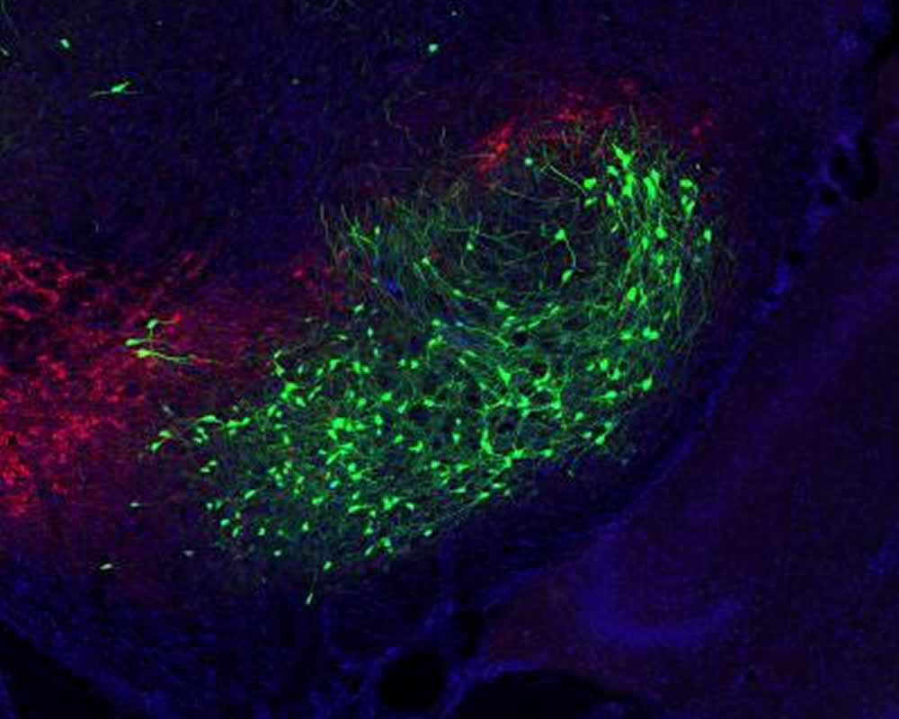
The scientists sought to trace these circuits in rodent models to better understand each step of neuronal communication. They focused on the emotion and movement brain loops, starting from a region involved in emotion (the medial prefrontal cortex) and a region involved in movement (the primary motor cortex). They used multiple genetic and viral tracing tools, including a technique developed by Salk Professor Ed Callaway’s lab, to observe how each loop was organized in the brain.
To the researchers’ surprise, they found a one-way communication pathway from the emotion loop to the movement loop through an area located deep in the brain called the basal ganglia. The basal ganglia, which includes structures involved in guiding behavior, essentially acts as a crossroads for the emotion circuit to directly influence the movement circuit to control action. To confirm their results, the authors used optogenetics, a technique that uses light to control cells, to investigate the precise function of the neurons in this region. They also studied each neural circuit in isolation from the rest of the brain and confirmed the novel pathway.
“Psychiatric diseases such as depression and anxiety can alter actions in a dramatic way by either decreasing or increasing movement. This mechanism represents a likely way that emotional states are related to changes in action control in psychiatric diseases,” says Jin.
Additionally, this unidirectional communication may be relevant for recovery from spinal cord injury. Researchers previously focused on movement centers of the brain because spinal cord injury is a movement issue; however, since these results suggest emotional states can influence brain movement centers, experiencing positive emotions such as motivation may aid patients in the recovery process. Activating emotion centers could likewise stimulate movement centers and facilitate recovery, according to the Salk co-first authors Jared Smith, a postdoctoral fellow, and Hao Li, a senior research associate. Further, these results suggest that emotional states could directly influence sports performance. So, Jin advices, maybe the next time you feel anxious during a game, just calm down and let the action take care of itself.
Other authors included Xunyi Yan of Salk; Masakazu Igarashi and Jeffery R. Wickens of the Okinawa Institute of Science and Technology; Patrice Coulon of the Centre National de la Recherche Scientifique (CNRS) and Aix-Marseille Université; and Tom J.H. Ruigrok of the Erasmus Medical Center Rotterdam.
Funding: The work was funded by the JSPS Institutional Program for Young Researcher Overseas Visits; the JSPS Grant-in-Aid for Young Scientist; the JSPS Grant-in-Aid for Challenging Exploratory Research; the JSPS Grant-in-Aid for JSPS Fellows; the Dutch Ministry of Health, Welfare, and Sports; the CNRS and Aix-Marseille Université through UMR 7289; the Human Frontier Science Program; the US National Institutes of Health (R01NS083815, R01AG047669, and K99NS106528 460); the Rose Hills Foundation and the McKnight Memory and Cognitive Disorders Award.
Source:
Salk Institute
Media Contacts:
Salk Communications – Salk Institute
Image Source:
The image is credited to Salk Institute.
Original Research: Open access
“An open cortico-basal ganglia loop allows limbic control over motor output via the nigrothalamic pathway”. Sho Aoki, Jared B Smith, Hao Li, Xunyi Yan, Masakazu Igarashi, Patrice Coulon, Jeffery R Wickens, Tom JH Ruigrok, Xin Jin.
eLife doi:10.7554/eLife.49995
Abstract
An open cortico-basal ganglia loop allows limbic control over motor output via the nigrothalamic pathway
Cortico-basal ganglia-thalamocortical loops are largely conceived as parallel circuits that process limbic, associative, and sensorimotor information separately. Whether and how these functionally distinct loops interact remains unclear. Combining genetic and viral approaches, we systemically mapped the limbic and motor cortico-basal ganglia-thalamocortical loops in rodents. Despite largely closed loops within each functional domain, we discovered a unidirectional influence of the limbic over the motor loop via ventral striatum-substantia nigra (SNr)-motor thalamus circuitry. Slice electrophysiology verifies that the projection from ventral striatum functionally inhibits nigro-thalamic SNr neurons. In vivo optogenetic stimulation of ventral or dorsolateral striatum to SNr pathway modulates activity in medial prefrontal cortex (mPFC) and motor cortex (M1), respectively. However, whereas the dorsolateral striatum-SNr pathway exerts little impact on mPFC, activation of the ventral striatum-SNr pathway effectively alters M1 activity. These results demonstrate an open cortico-basal ganglia loop whereby limbic information could modulate motor output through ventral striatum control of M1.


