Summary: A new study sheds light on how the brain processes rewards and provides a deeper understanding on the neurobiology of depression.
Source: University of Maryland School of Medicine.
In new pre-clinical research, scientists at the University of Maryland School of Medicine (UMSOM), led by Scott Thompson, PhD, Professor of Physiology, have identified changes in brain activity linked to the pleasure and reward system.
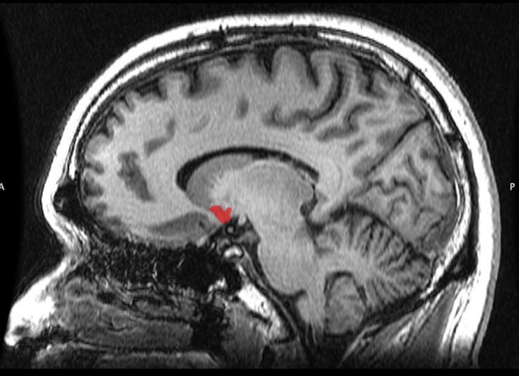
The research, published in the journal, Nature, provides news insights into how the brain processes rewards, and advances our understanding of addiction and depression. The research, which was conducted by Tara LeGates, Ph.D., a Research Associate in the Department of Physiology, discovered that the strength of signals between two brain regions, the hippocampus and the nucleus accumbens, is critical for processing information related to a rewarding stimulus, such as its location.
“These two parts of the brain are known to be important in processing rewarding experiences,” said Dr. Thompson.” The communication between these regions is stronger in addiction, although the mechanisms underlying this were unknown. We also suspected that opposite changes in the strength of this communication would occur in depression. A weakening of their connections could explain the defect in reward processing that causes the symptom of anhedonia in depressed patients.” Anhedonia is the inability to enjoy normally pleasurable experiences, such as food, being with friends or family, and sex.
This research uncovered a key circuit in the brain of mice that is important for goal-directed behaviors and shows that the strength of the signals in this circuit are changeable, a process referred to as plasticity. Reward circuits and the molecular components that underlie their plasticity represent new targets for development of treatments for disorders like addiction and depression.
Using Light-Sensitive Proteins
To activate or inhibit this connection, researchers used special light-sensitive proteins introduced into specific neurons in the brains of the mice. In mice with the light-sensitive protein that stimulated the neurons, just four seconds of light exposure not only activated this hippocampus-to-nucleus accumbens pathway while the light was on but persistently reinforced the strength of the signals along this pathway, creating an artificial reward memory.
A day later, the mice returned to the place where the artificial memory was created, even though they never experienced an actual reward there. Researchers then used light to silence the same pathway in mice with the light-sensitive protein that inhibited the neurons and found that this pathway is required for associating a reward with its location. The mice no longer showed a preference for the place where they had interacted with another mouse.

The researchers also examined this circuit in depressed mice. This pathway could not be enhanced using the stimulating light-sensitive protein. After receiving anti-depressant medication, the researchers could enhance this pathway using the light-sensitive protein and create artificial reward memories in the mice.
This work was funded by the National Institutes of Health, the Whitehall Foundation, and the Brain & Behavior Research Foundation (NARSAD Young Investigator Grant).
“These exciting results bring us closer to understanding what goes wrong in the brains of clinically depressed patients,” said UMSOM Dean E. Albert Reece, MD, PhD, MBA, who is also the Executive Vice President for Medical Affairs, University of Maryland, and the John Z. and Akiko K. Bowers Distinguished Professor.
Funding: This research was funded by National Institutes of Health, Whitehall Foundation, Brain & Behavior Research Foundation.
Source: Joanne Morrison – University of Maryland School of Medicine
Publisher: Organized by NeuroscienceNews.com.
Image Source: NeuroscienceNews.com image is in the public domain.
Original Research: Abstract for “Reward behaviour is regulated by the strength of hippocampus–nucleus accumbens synapses” by Tara A. LeGates, Mark D. Kvarta, Jessica R. Tooley, T. Chase Francis, Mary Kay Lobo, Meaghan C. Creed & Scott M. Thompson in Nature. Published November 26 2018.
doi:10.1038/s41586-018-0740-8
[cbtabs][cbtab title=”MLA”]University of Maryland School of Medicine”Clues to Brain Changes in Depression Discovered.” NeuroscienceNews. NeuroscienceNews, 27 November 2018.
<https://neurosciencenews.com/depression-brain-changes-10263/>.[/cbtab][cbtab title=”APA”]University of Maryland School of Medicine(2018, November 27). Clues to Brain Changes in Depression Discovered. NeuroscienceNews. Retrieved November 27, 2018 from https://neurosciencenews.com/depression-brain-changes-10263/[/cbtab][cbtab title=”Chicago”]University of Maryland School of Medicine”Clues to Brain Changes in Depression Discovered.” https://neurosciencenews.com/depression-brain-changes-10263/ (accessed November 27, 2018).[/cbtab][/cbtabs]
Abstract
Reward behaviour is regulated by the strength of hippocampus–nucleus accumbens synapses
Reward drives motivated behaviours and is essential for survival, and therefore there is strong evolutionary pressure to retain contextual information about rewarding stimuli. This drive may be abnormally strong, such as in addiction, or weak, such as in depression, in which anhedonia (loss of pleasure in response to rewarding stimuli) is a prominent symptom. Hippocampal input to the shell of the nucleus accumbens (NAc) is important for driving NAc activity1,2 and activity-dependent modulation of the strength of this input may contribute to the proper regulation of goal-directed behaviours. However, there have been few robust descriptions of the mechanisms that underlie the induction or expression of long-term potentiation (LTP) at these synapses, and there is, to our knowledge, no evidence about whether such plasticity contributes to reward-related behaviour. Here we show that high-frequency activity induces LTP at hippocampus–NAc synapses in mice via canonical, but dopamine-independent, mechanisms. The induction of LTP at this synapse in vivo drives conditioned place preference, and activity at this synapse is required for conditioned place preference in response to a natural reward. Conversely, chronic stress, which induces anhedonia, decreases the strength of this synapse and impairs LTP, whereas antidepressant treatment is accompanied by a reversal of these stress-induced changes. We conclude that hippocampus–NAc synapses show activity-dependent plasticity and suggest that their strength may be critical for contextual reward behaviour.


