

Summary: Combining infrared laser stimulation with ultra-high-field MRI, researchers revolutionize brain mapping.
Source: Oregon Health and Science University
Scientists have discovered a new method for quickly and efficiently mapping the vast network of connections among neurons in the brain.
Researchers combined infrared laser stimulation techniques with functional magnetic resonance imaging in animals to generate mapping of connections throughout the brain. The technique was described in a study published in the journal Science Advances.
“This is a revolution in detecting connections in the brain,” said senior author Anna Wang Roe, Ph.D., a professor in the Division of Neuroscience at OHSU’s Oregon National Primate Research Center. “The ability to easily map connections in the living brain with high precision opens doors for other applications in medicine and engineering.”
Roe led an international team of researchers in the United States and China. Roe splits her time between her lab at OHSU in Portland, Oregon, and Zhejiang University in Hangzhou, China, where she directs the Interdisciplinary Institute of Neuroscience and Technology.
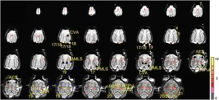
Researchers threaded a 200-micron optical fiber into the brains of research animals – in this case, cats and monkeys at Zhejiang and Vanderbilt, respectively – and stimulated specific areas of the brain. They were then able to view the cascading series of connections through ultra-high-field MRIs measuring blood oxygen levels in various areas.
Traditionally, researchers have mapped these kinds of connections in the brain through a laborious process in which they injected dyes directly into the brain and reconstructed the connections post-mortem.
“It’s a very slow, expensive and time-consuming process,” she said.
The new technique opens doors for systematic, large-scale study of connection patterns within single individuals repeatedly and efficiently. It also enables researchers to determine the direction of information flowing within the brain, an insight critical for understanding information processing in the brain.
Roe said she believes the technique will greatly expand scientists’ ability to understand the brain and could accelerate development of artificial intelligence and technologies that use brain-machine interface.
“If we can understand the organizing patterns in the brain, this could lead to some understanding of the general rules of connectivity,” she said.
Funding: Funding for this research was supported by the National Natural Science Foundation, grants 81430010 and 31627802, 81701774, 61771423 and 31471052, National Hi-Tech Research and Development Program grant 205AA020515, Zhejiang Provincial Natural Science Foundation of China grant LR15C090001, Fundamental Research Funds for the Central Universities grants 2015QN81007 and 2016QN81018, National Institutes of Health grants N5044375 and NS052407.
Source:
Oregon Health and Science University
Media Contacts:
Erik Robinson – Oregon Health and Science University
Image Source:
The image is Anna Wang Roe et al./Science Advances.
Original Research: Open access
“Focal infrared neural stimulation with high-field functional MRI: A rapid way to map mesoscale brain connectomes”. Augix Guohua Xu, Meizhen Qian, Feiyan Tian, Bin Xu, Robert M. Friedman, Jianbao Wang, Xuemei Song, Yi Sun, Mykyta M. Chernov, Jonathan M. Cayce, E. Duco Jansen, Anita Mahadevan-Jansen, Xiaotong Zhang, Gang Chen and Anna Wang Roe.
Science Advances 24 APR 2019 doi:10.1126/sciadv.aau7046
Abstract
Focal infrared neural stimulation with high-field functional MRI: A rapid way to map mesoscale brain connectomes
We have developed a way to map brain-wide networks using focal pulsed infrared neural stimulation in ultrahigh-field magnetic resonance imaging (MRI). The patterns of connections revealed are similar to those of connections previously mapped with anatomical tract tracing methods. These include connections between cortex and subcortical locations and long-range cortico-cortical connections. Studies of local cortical connections reveal columnar-sized laminar activation, consistent with feed-forward and feedback projection signatures. This method is broadly applicable and can be applied to multiple areas of the brain in different species and across different MRI platforms. Systematic point-by-point application of this method may lead to fundamental advances in our understanding of brain connectomes.


