

Summary: Mice infected with bacteria that cause mild intestinal infections exhibited Parkinson’s like symptoms later in life. The findings provide a pathophysiological model in which intestinal infections may act as a trigger for Parkinson’s disease.
Source: University of Montreal
A new study by Montreal scientists published today in Nature demonstrates that a gut infection can lead to a pathology resembling Parkinson’s disease (PD) in a mouse model lacking a gene linked to the human disease.
This discovery extends recent work by the same group suggesting that PD has a major immune component, providing new avenues for therapeutic strategies.
The collaborative study was the work of a joint team of scientists led by Michel Desjardins and Louis-Eric Trudeau of Université de Montréal, Heidi McBride at the Montreal Neurological Institute, and Samantha Gruenheid at McGill University.
The number of PD patients in the world more than doubled between 1990 and 2016, from 2.5 million to 6.1 million. A relatively conservative projected doubling of the number of patients over the next 30 years would yield more than 12 million patients worldwide by about 2050.
About 10 percent of cases of PD are due to mutations in genes coding for proteins such as PINK1 and Parkin, which have been linked to mitochondria (the organelle in cells that produces energy). Patients with these mutations develop PD at a much earlier age. However, in mouse models, the same mutations do not generate disease symptoms, leading many researchers to conclude that mice may not be suitable for the study of PD.
Trudeau and McBride, two specialists in the field of PD research, argue that the findings in this new study may explain this disparity: these animals are normally kept in germ-free facilities, conditions not representative of those encountered by human beings who are constantly exposed to infectious microorganisms. Gruenheid, a microbiologist, is confident that the link between infection and PD will stimulate further study of the immune response linked to the initiation of the disease, allowing researchers to develop and test novel therapeutic approaches.
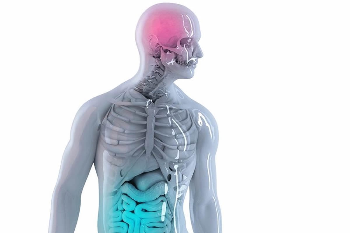
PD is caused by the progressive death of a subset of neurons in the brain, called dopaminergic neurons. This loss of neurons is responsible for the typical motor symptoms observed in PD patients, including tremors and rigidity. What causes the death of the dopaminergic neurons is still unknown.
“Most of the current models of PD are based on the belief that neurons die due to toxic elements accumulating inside them,” said Trudeau, a neuroscientist.
“This does not explain, however, the fact that PD pathology is initiated in patients several years before the emergence of the motor impairment and any noticeable loss of neurons.”
The reason for this is believed to be explained by the results of the present study. The Montreal team has shown that in mice lacking a gene linked to PD, infection with bacteria that cause mild intestinal symptoms in young mice was sufficient to trigger PD-like symptoms in these animals later in life.
The Parkinson’s-like symptoms could be temporarily reversed by the administration of L-DOPA, a drug used to treat Parkinson’s patients. Desjardins and Diana Matheoud, an immunologist the University of Montreal Hospital Research Centre (CRCHUM), point out that in normal mice, the immune system responded properly to the gut infection; however, in mice lacking the gene PINK1 related to Parkinson’s, the immune system overreacted and triggered “auto-immunity”, a process that leads the immune system to attack healthy cells in the organism. The results published today suggest that rather than dying from toxin accumulation, the killing of dopaminergic neurons involves immune cells.
In the infected mutant mice, autoreactive toxic T lymphocytes were shown to be present in the brain and able to attack healthy neurons in culture dishes. The co-first authors of the published article, Matheoud and Tyler Cannon, a graduate student in microbiology, emphasize the fact that these results strongly suggest that some forms of PD are an autoimmune disease likely to start in the gut several years before patients notice any motor symptoms, highlighting the fact that a window of time exists for preventive treatment.
This exciting collaborative discovery required the expertise of a microbiologist, immunologist, neuroscientist, and a cell biologist, highlighting the complexity of this devastating disease.
Funding: Support for the work was provided through the Canadian Institutes of Health Research, the Brain Canada and Krembil Foundations and The Michael J. Fox Foundation for Parkinson’s Research.
Source:
University of Montreal
Media Contacts:
Genevieve O’Meara – University of Montreal
Image Source:
The image is adapted from the University of Montreal news release.
Original Research: Closed access
“Intestinal infection triggers Parkinson’s disease-like symptoms in Pink1−/− mice”. Diana Matheoud, Tyler Cannon, Aurore Voisin, Anna-Maija Penttinen, Lauriane Ramet, Ahmed M. Fahmy, Charles Ducrot, Annie Laplante, Marie-Josée Bourque, Lei Zhu, Romain Cayrol, Armelle Le Campion, Heidi M. McBride, Samantha Gruenheid, Louis-Eric Trudeau & Michel Desjardin.
Nature. doi:10.1038/s41586-019-1405-y
Abstract
Intestinal infection triggers Parkinson’s disease-like symptoms in Pink1−/− mice
Parkinson’s disease is a neurodegenerative disorder with motor symptoms linked to the loss of dopaminergic neurons in the substantia nigra compacta. Although the mechanisms that trigger the loss of dopaminergic neurons are unclear, mitochondrial dysfunction and inflammation are thought to have key roles. An early-onset form of Parkinson’s disease is associated with mutations in the PINK1 kinase and PRKN ubiquitin ligase genes. PINK1 and Parkin (encoded by PRKN) are involved in the clearance of damaged mitochondria in cultured cells, but recent evidence obtained using knockout and knockin mouse models have led to contradictory results regarding the contributions of PINK1 and Parkin to mitophagy in vivo. It has previously been shown that PINK1 and Parkin have a key role in adaptive immunity by repressing presentation of mitochondrial antigens, which suggests that autoimmune mechanisms participate in the aetiology of Parkinson’s disease. Here we show that intestinal infection with Gram-negative bacteria in Pink1−/− mice engages mitochondrial antigen presentation and autoimmune mechanisms that elicit the establishment of cytotoxic mitochondria-specific CD8+ T cells in the periphery and in the brain. Notably, these mice show a sharp decrease in the density of dopaminergic axonal varicosities in the striatum and are affected by motor impairment that is reversed after treatment with L-DOPA. These data support the idea that PINK1 is a repressor of the immune system, and provide a pathophysiological model in which intestinal infection acts as a triggering event in Parkinson’s disease, which highlights the relevance of the gut–brain axis in the disease.


