

Summary: Convolutional neural network model significantly outperforms previous methods and is as accurate as humans in segmenting active and overlapping neurons.
Source: Duke University
Biomedical engineers at Duke University have developed an automated process that can trace the shapes of active neurons as accurately as human researchers can, but in a fraction of the time.
This new technique, based on using artificial intelligence to interpret video images, addresses a critical roadblock in neuron analysis, allowing researchers to rapidly gather and process neuronal signals for real-time behavioral studies.
The research appeared this week in the Proceedings of the National Academy of Sciences.
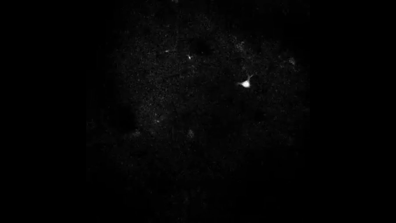
To measure neural activity, researchers typically use a process known as two-photon calcium imaging, which allows them to record the activity of individual neurons in the brains of live animals. These recordings enable researchers to track which neurons are firing, and how they potentially correspond to different behaviors.
While these measurements are useful for behavioral studies, identifying individual neurons in the recordings is a painstaking process. Currently, the most accurate method requires a human analyst to circle every ‘spark’ they see in the recording, often requiring them to stop and rewind the video until the targeted neurons are identified and saved. To further complicate the process, investigators are often interested in identifying only a small subset of active neurons that overlap in different layers within the thousands of neurons that are imaged.
This process, called segmentation, is fussy and slow. A researcher can spend anywhere from four to 24 hours segmenting neurons in a 30-minute video recording, and that’s assuming they’re fully focused for the duration and don’t take breaks to sleep, eat or use the bathroom.
In contrast, a new open source automated algorithm developed by image processing and neuroscience researchers in Duke’s Department of Biomedical Engineering can accurately identify and segment neurons in minutes.
“As a critical step towards complete mapping of brain activity, we were tasked with the formidable challenge of developing a fast automated algorithm that is as accurate as humans for segmenting a variety of active neurons imaged under different experimental settings,” said Sina Farsiu, the Paul Ruffin Scarborough Associate Professor of Engineering in Duke BME.
“The data analysis bottleneck has existed in neuroscience for a long time — data analysts have spent hours and hours processing minutes of data, but this algorithm can process a 30-minute video in 20 to 30 minutes,” said Yiyang Gong, an assistant professor in Duke BME. “We were also able to generalize its performance, so it can operate equally well if we need to segment neurons from another layer of the brain with different neuron size or densities.”
“Our deep learning-based algorithm is fast, and is demonstrated to be as accurate as (if not better than) human experts in segmenting active and overlapping neurons from two-photon microscopy recordings,” said Somayyeh Soltanian-Zadeh, a PhD student in Duke BME and first author on the paper.
Deep-learning algorithms allow researchers to quickly process large amounts of data by sending it through multiple layers of nonlinear processing units, which can be trained to identify different parts of a complex image. In their framework, this team created an algorithm that could process both spatial and timing information in the input videos. They then ‘trained’ the algorithm to mimic the segmentation of a human analyst while improving the accuracy.
The advance is a critical step towards allowing neuroscientists to track neural activity in real time. Because of their tool’s widespread usefulness, the team has made their software and annotated dataset available online.
Gong is already using the new method to more closely study the neural activity associated with different behaviors in mice. By better understanding which neurons fire for different activities, Gong hopes to learn how researchers can manipulate brain activity to modify behavior.
“This improved performance in active neuron detection should provide more information about the neural network and behavioral states, and open the door for accelerated progress in neuroscience experiments,” said Soltanian-Zadeh.
Funding: This work was supported by the National Institutes of Health Medical Imaging Training Program pre-doctoral fellowship (T32-EB001040 and P30-EY005722) and the National Science Foundation BRAIN Initiative (NCS-FO 1533598).Yiyang Gong is supported by the Beckman Young Investigator Award, and Sina Farsiu is supported by a Google Faculty Research Award.
Source:
Duke University
Media Contacts:
Sina Farsiu – Duke University
Image Source:
The image is credited to Yiyang Gong, Duke University.
Original Research: Closed access.
“Fast and Robust Active Neuron Segmentation in Two-Photon Calcium Imaging Using Spatio-Temporal Deep-Learning,” Somayyeh Soltanian-Zadeh, Kaan Sahingur, Sarah Blau, Yiyang Gong, and Sina Farsiu. Proceedings of the National Academy of Sciences, April 12, 2019. doi:10.1073/pnas.1812995116
Abstract
Fast and robust active neuron segmentation in two-photon calcium imaging using spatiotemporal deep learning
Calcium imaging records large-scale neuronal activity with cellular resolution in vivo. Automated, fast, and reliable active neuron segmentation is a critical step in the analysis workflow of utilizing neuronal signals in real-time behavioral studies for discovery of neuronal coding properties. Here, to exploit the full spatiotemporal information in two-photon calcium imaging movies, we propose a 3D convolutional neural network to identify and segment active neurons. By utilizing a variety of two-photon microscopy datasets, we show that our method outperforms state-of-the-art techniques and is on a par with manual segmentation. Furthermore, we demonstrate that the network trained on data recorded at a specific cortical layer can be used to accurately segment active neurons from another layer with different neuron density. Finally, our work documents significant tabulation flaws in one of the most cited and active online scientific challenges in neuron segmentation. As our computationally fast method is an invaluable tool for a large spectrum of real-time optogenetic experiments, we have made our open-source software and carefully annotated dataset freely available online.


