The visual system keeps track of relevant objects even as eye movements are made, shows a study by the German Primate Center.
When watching basketball, we are easily able to keep track of the ball while also making frequent eye and head movements to look at the different players. Neuroscientists Tao Yao, Stefan Treue and B. Suresh Krishna from the German Primate Center (DPZ) in Göttingen, Germany, wanted to understand the neural mechanisms that allow us to see a stable world and keep track of relevant objects even without directly looking at them and when we shift our gaze. Their study shows that the rhesus macaque’s brain “marks” relevant visual objects and rapidly updates the position of these markers as the monkey looks around. Since humans and monkeys exhibit very similar eye-movements and visual function, these findings are likely to generalize to the human brain. These results are also likely to be important for our understanding of disorders like schizophrenia, visual neglect and other attention deficit disorders.
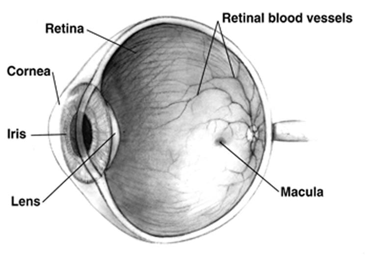
The light that enters the eye falls onto the retina, where it is converted into neural activity that is then used by the brain to provide our sense of vision. The central part of the retina, the fovea, is specialized for more sensitive, higher-definition vision. It is therefore advantageous when viewing a scene, to move the eye so that it is centered successively (or fixated) on each important part of the scene, and light from these parts can fall onto the fovea and be analyzed in greater detail. Indeed, both humans and monkeys make two to three fast eye movements every second in this manner, with each eye-movement lasting less than one-tenth of a second. Because the eye acts like a camera, each eye-movement results in a different view of the scene falling onto the retina.
However, despite these fast changes in viewpoint (which can also result from head movements), humans and monkeys do not see a scene that jumps around: Instead, they are able to “stitch together” the information obtained during each fixation to perceive a stable visual scene. They are also able to keep track of where relevant objects are in the scene even with these frequent changes in viewpoint. This is a very challenging task. Visual neurons respond more to relevant objects than to irrelevant ones. This increased response to relevant stimuli “marks” relevant stimuli. Since each visual neuron in the brain only responds when a specific part of the retina is stimulated, each change in viewpoint with an eye-movement results in a different group of neurons being activated by a given visual stimulus before and after the eye-movement. This means that the “marking”, i.e. the information about which objects are relevant, needs to be transferred between different groups of neurons, so that after the eye-movement, these relevant objects continue to evoke larger responses and the brain can keep track of them. However, very little was known about the properties of such an information transfer in the brain, or even about whether it occurred at all.

In order to address this, neuroscientists Tao Yao, Stefan Treue and Suresh Krishna of the German Primate Center (DPZ) examined the responses of many single neurons in the brain of two monkeys while they attended to a stimulus without directly looking at it and made an eye-movement while maintaining attention on this stimulus. To measure the activity of single neurons, the scientists inserted electrodes thinner than a human hair into the monkey’s brain and recorded the neurons’ electrical activity. Because the brain is not pain-sensitive, this insertion of electrodes is painless for the animal. By recording from single neurons in an area of the monkey’s brain known as area MT, the scientists were able to show that a transfer of information about the locations of relevant objects indeed occurs. However, no information is transferred about what the relevant objects look like.
“Our study shows how the primate brain is able to keep track of attended objects while ignoring irrelevant ones”, says Tao Yao, first author of the publication. It supports the idea that the brain maintains markers to attended stimuli and updates the locations of these markers with each eye-movement. “Our results answer several important questions about how our brains see a stable visual world despite frequent intervening eye-movements. Also, because the updating of attentional markers is known to be impaired in schizophrenia, visual neglect and other attention deficit disorders, our results may help improve our understanding of these diseases”, Tao Yao comments on the findings.
NeuroscienceNews.com could like to thank Susanne Diederich for submitting this research news directly to us.
Source: Susanne Diederich – DPZ
Image Source: The image is in the public domain.
Original Research: Full open access research for “An attention-sensitive memory trace in macaque MT following saccadic eye movements” by Tao Yao, Stefan Treue and B. Suresh Krishna in PLOS Biology. Published online February 22 2016 doi:10.1371/journal.pbio.1002390
Abstract
Rehabilitation drives enhancement of neuronal structure in functionally relevant neuronal subsets
We experience a visually stable world despite frequent retinal image displacements induced by eye, head, and body movements. The neural mechanisms underlying this remain unclear. One mechanism that may contribute is transsaccadic remapping, in which the responses of some neurons in various attentional, oculomotor, and visual brain areas appear to anticipate the consequences of saccades. The functional role of transsaccadic remapping is actively debated, and many of its key properties remain unknown. Here, recording from two monkeys trained to make a saccade while directing attention to one of two spatial locations, we show that neurons in the middle temporal area (MT), a key locus in the motion-processing pathway of humans and macaques, show a form of transsaccadic remapping called a memory trace. The memory trace in MT neurons is enhanced by the allocation of top-down spatial attention. Our data provide the first demonstration, to our knowledge, of the influence of top-down attention on the memory trace anywhere in the brain. We find evidence only for a small and transient effect of motion direction on the memory trace (and in only one of two monkeys), arguing against a role for MT in the theoretically critical yet empirically contentious phenomenon of spatiotopic feature-comparison and adaptation transfer across saccades. Our data support the hypothesis that transsaccadic remapping represents the shift of attentional pointers in a retinotopic map, so that relevant locations can be tracked and rapidly processed across saccades. Our results resolve important issues concerning the perisaccadic representation of visual stimuli in the dorsal stream and demonstrate a significant role for top-down attention in modulating this representation.
“An attention-sensitive memory trace in macaque MT following saccadic eye movements” by Tao Yao, Stefan Treue and B. Suresh Krishna in PLOS Biology. Published online February 22 2016 doi:10.1371/journal.pbio.1002390


