

Summary: A new study reports on how ischemic stroke can cause long term damage to the blood-spinal cord barrier.
Source: USF Health.
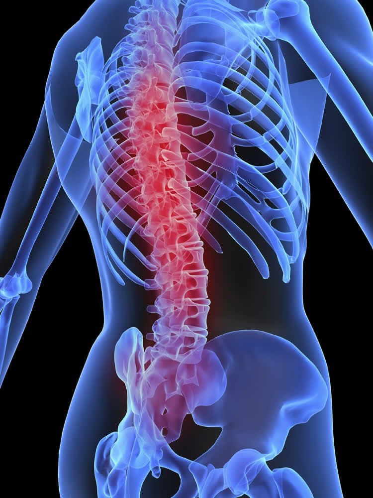
Stroke’s long-term effects on blood-spinal cord barrier can lead to ‘an increasingly toxic environment’ in spinal cord and ‘significant input on disease pathology’.
A team of researchers at the University of South Florida investigating the short and long-term effects of ischemic stroke in a rodent model has found that stroke can cause long-term damage to the blood-spinal cord barrier (BSCB), creating a “toxic environment” in the spinal cord that might leave stroke survivors susceptible to motor dysfunction and disease pathology.
The paper describing their study was recently published online and will appear in an upcoming issue of Journal of Neuropathology and Experimental Neurology.
“This study, carried out using laboratory rats modeling stroke, demonstrated that ischemic stroke – in both its subacute and chronic stages – damages the BSCB in a variety of ways, creating a toxic environment in the spinal cord that can lead to further disability and exacerbate disease pathology,” said study lead author Dr. Svitlana Garbuzova-Davis, associate professor in USF’s Center of Excellence for Aging and Brain Repair, Department of Neurosurgery and Brain Repair. “The aim of our study was to evaluate post-stroke BSCB condition that might lead to the development of more effective therapies for stroke survivors.”
The BSCB provides a specialized protective ‘microenvironment’ for neural cells in the spinal cord. Substantial vascular damage is a major pathologic feature of both subacute and chronic stroke caused by an extended period of microvascular permeability after the BSCB loses integrity. Damage to the BSCB, explained the researchers, plays a fundamental role in the development of several pathological conditions, including abnormal motor function.
The researchers, who evaluated the BSCB in test animals at seven and 30 days after stroke modeling, found that ischemic stroke damaged the gray and white matter in the cervical spinal cord on both sides of the spinal column, based on analysis of electron microscope images. Among the effects were damage to neural cells called ‘astrocytes,’ loss of motor neurons, reduced integrity of a tight junction protein between barrier cells, and swollen axons with damaged myelin in ascending and descending tracts connecting to the brain.
They also found stroke-associated ‘upregulation’ of Beclin-1 in endothelial cells composing the BSCB. Beclin-1, explained the researchers, helps induce autophagy, an activity associated with removal of various intracellular components. They also observed a decrease in LC3B, an essential autophagy protein, at a later stage post-stroke. These observations of Beclin-1 and LC3B suggest an impaired post-stroke autophagy process in spinal cord capillaries, inducing endothelial cell degeneration.
These stroke-related alterations in the cervical spinal cord indicate pervasive and long-lasting BSCB damage that would severely affect spinal cord function, wrote the researchers, adding that the widespread microvascular impairment in the gray and white matter of the cervical spinal cord aggravated motor neuron deterioration and had the potential to cause motor dysfunction.
“Because our investigations on the post-stroke microvascular alterations, including BSCB damage, have just begun, many questions remain,” said senior author Dr. Cesario Borlongan, professor and director of the USF Center of Excellence for Aging and Brain Repair. “Specifically, the protein expression responsible for endothelial cell degeneration and tight junction damage we identified in this study needs to be confirmed through further tests. Also, behavioral tests of motor function in post-stroke animals in correlation with BSCB damage are needed. These questions and others will be addressed in our future studies.”
Dr. Paul R. Sanberg, Distinguished University Professor, a co-author of the paper, concluded that “these novel data showing BSCB damage in subacute and chronic ischemic stroke may lead to development of new therapeutic approaches for patients with ischemic cerebral infarction.”
Source: Svitlana Garbuzova-Davis – USF Health
Image Source: This NeuroscienceNews.com image is in the public domain.
Original Research: Abstract for “Blood-Spinal Cord Barrier Alterations in Subacute and Chronic Stages of a Rat Model of Focal Cerebral Ischemia” by Svitlana Garbuzova-Davis, Edward Haller, Naoki Tajiri, Avery Thomson, Jennifer Barretta, Stephanie N. Williams, Eithan D. Haim, Hua Qin, Aric Frisina-Deyo, Jerry V. Abraham, Paul R. Sanberg, Harry Van Loveren, and Cesario V. Borlongan in Journal of Neuropathology & Experimental Neurology. Published online June 9 2016 doi:10.1093/jnen/nlw040
[cbtabs][cbtab title=”MLA”]USF Health. “Strokes Damage the Blood-Spinal Cord Barrier.” NeuroscienceNews. NeuroscienceNews, 13 June 2016.
<https://neurosciencenews.com/stroke-blood-spinal-cord-barrier-4458/>.[/cbtab][cbtab title=”APA”]USF Health. (2016, June 13). Strokes Damage the Blood-Spinal Cord Barrier. NeuroscienceNews. Retrieved June 13, 2016 from https://neurosciencenews.com/stroke-blood-spinal-cord-barrier-4458/[/cbtab][cbtab title=”Chicago”]USF Health. “Strokes Damage the Blood-Spinal Cord Barrier.” https://neurosciencenews.com/stroke-blood-spinal-cord-barrier-4458/ (accessed June 13, 2016).[/cbtab][/cbtabs]
Abstract
Blood-Spinal Cord Barrier Alterations in Subacute and Chronic Stages of a Rat Model of Focal Cerebral Ischemia
We previously demonstrated blood-brain barrier impairment in remote contralateral brain areas in rats at 7 and 30 days after transient middle cerebral artery occlusion (tMCAO), indicating ischemic diaschisis. Here, we focused on effects of subacute and chronic focal cerebral ischemia on the blood-spinal cord barrier (BSCB). We observed BSCB damage on both sides of the cervical spinal cord in rats at 7 and 30 days post-tMCAO. Major BSCB ultrastructural changes in spinal cord gray and white matter included vacuolated endothelial cells containing autophagosomes, pericyte degeneration with enlarged mitochondria, astrocyte end-feet degeneration and perivascular edema; damaged motor neurons, swollen axons with unraveled myelin in ascending and descending tracts and astrogliosis were also observed. Evans Blue dye extravasation was maximal at 7 days. There was immunofluorescence evidence of reduction of microvascular expression of tight junction occludin, upregulation of Beclin-1 and LC3B immunoreactivities at 7 days and a reduction of the latter at 30 days post-ischemia. These novel pathological alterations on the cervical spinal cord microvasculature in rats after tMCAO suggest pervasive and long-lasting BSCB damage after focal cerebral ischemia, and that spinal cord ischemic diaschisis should be considered in the pathophysiology and therapeutic approaches in patients with ischemic cerebral infarction.
“Blood-Spinal Cord Barrier Alterations in Subacute and Chronic Stages of a Rat Model of Focal Cerebral Ischemia” by Svitlana Garbuzova-Davis, Edward Haller, Naoki Tajiri, Avery Thomson, Jennifer Barretta, Stephanie N. Williams, Eithan D. Haim, Hua Qin, Aric Frisina-Deyo, Jerry V. Abraham, Paul R. Sanberg, Harry Van Loveren, and Cesario V. Borlongan in Journal of Neuropathology & Experimental Neurology. Published online June 9 2016 doi:10.1093/jnen/nlw040


