

It is widely recognised that our physical fitness is reflected in our mental fitness, especially as we get older. How does being physically fit affect our aging brains? Neuroimaging studies, in which the activity of different parts of the brain can be visualised, have provided some clues. Until now, however, no study has directly linked brain activation with both mental and physical performance.
As reported in the latest volume of the journal NeuroImage, an exciting new study led by Dr Hideaki Soya from the University of Tsukuba in Japan and his colleagues show, for the first time, the direct relationship between brain activity, brain function and physical fitness in a group of older Japanese men. They found that the fitter men performed better mentally than the less fit men, by using parts of their brains in the same way as in their youth.
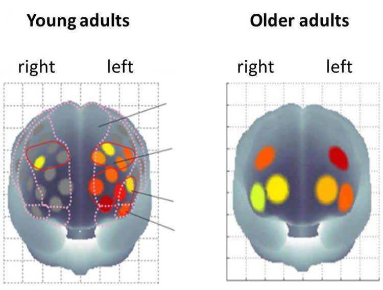
As we age, we use different parts of our brain compared to our younger selves. For example, when young, we mainly use the left side of our prefrontal cortex (PFC) for mental tasks involving short term memory, understanding the meaning of words and the ability to recognize previously encountered events, objects, or people. When older, we tend to use the equivalent parts of our PFC on the right side of the brain for these tasks. The PFC is located in the very front of the brain, just behind the forehead. It has roles in executive function, memory, intelligence, language and vision.
With tasks involving the temporary storage and manipulation of memory, long term memories and inhibitory control, young adults favor the right side of the PFC, while older adults engage both the right and left PFC. In fact, with aging, we tend to use both sides of
the PFC during mental tasks, rather than just one. This phenomenon has been coined HAROLD (hemispheric asymmetry reduction in older adults) and reflects the reorganisation of the brain as compensation for reduced brain capacity and efficiency due to age-related structural and physiological decline.
In the NeuroImage study, 60 older men (aged 64-75 years) underwent an exercise test to measure their aerobic fitness. The men, whose physical fitness was found to vary widely, then performed a test to measure their selective attention, executive function and reaction time. This well-known ‘color-word matching Stroop test’ involved showing the men words meaning color, such as blue, green, red, but asking them to name the color of the letters rather than read the word itself. This is harder than it sounds. When the color of the letters does not match the word – blue, red, green – it takes the brain longer to react. This reaction time is used as a measurement of brain function. Activity in the PFC region of the mens’ brains was measured throughout the test using a unique neuroimaging technique called functional near infrared spectroscopy or fNIRS. This technique provides a measure of blood oxygen concentration in surface blood vessels, indicative of activity in the brain’s outer layers, using a set of wearable probes in a cap that is placed on the head. Active brain cells require fresh oxygenated blood which dislodges the deoxygenated blood from that region. fNIRS measures the changes in color between oxygenated red blood and blue deoxygenated blood and thus indirectly measures brain activity.
The results from these tests were combined and extensively statistically analysed to explore the associations between aerobic fitness, Stroop reaction time and brain activity during the Stroop test. As predicted for older adults, during the Stroop test both sides of the PFC are active, with no difference between right and left, verifying the HAROLD phenomenon amongst this group of men. Previous studies have shown that young adults favour the left side of the PFC for this task.
Analysis of the relationship between brain activity and Stroop reaction time revealed that those men that favored the left side of the PFC while performing the Stroop test had faster reaction times. This indicates that older adults who use the more youth-like, task-related side of the brain perform better in this test.
Next, the association between aerobic fitness and Stroop reaction time was analysed. Fitter men had shorter reaction times.
Based on these findings, the researchers correctly predicted that higher aerobic fitness would be associated with higher left-PFC activity. In other words, fitter men tend to use the more youth-like side of their brains, at least while performing the Stroop test.
Previous studies have not examined the interaction between the three factors under investigation in this study – aerobic fitness, mental performance and brain activation. Using clever statistical tests called mediation analyses to look at these interactions, the researchers found that aerobically fitter older men can perform better mentally than less fit older men by using the more important brain regions when needed. In fact, the fitter older men are using parts of their brains in the same way as when they were younger.
How do they do this? Professor Soya says “one possible explanation suggested by the research is that the volume and integrity of the white matter in the part of brain that links the two sides declines with age. There is some evidence to support the theory that fitter adults are able to better maintain this white matter than less fit adults, but further study is needed to confirm this theory.”
If you are an aging woman, you will be wondering if these results can be applied to your female brain. Both aging sexes might also wonder whether increasing aerobic fitness later in life can increase mental fitness. The results aren’t in, but I’m heading off for a brisk walk just in case.
Source: University of Tsukuba
Image Source: The image is credited to the researchers/University of Tsukuba.
Original Research: Abstract for “The association between aerobic fitness and cognitive function in older men mediated by frontal lateralization” by Kazuki Hyodo, Ippeita Dan, Yasushi Kyutoku, Kazuya Suwabe, Kyeongho Byun, Genta Ochi, Morimasa Kato, and Hideaki Soya in NeuroImage. Published online October 9 2015 doi:10.1016/j.neuroimage.2015.09.062
Abstract
The association between aerobic fitness and cognitive function in older men mediated by frontal lateralization
Recent studies have gradually revealed that higher aerobic fitness is related to higher cognitive function and higher task-related prefrontal activation in older adults. However, a holistic picture of these factors has yet to be presented. As a typical age-related change of brain activation, less lateralized activity in the prefrontal cortex during cognitive tasks has been observed in various neuroimaging studies. Thus, this study aimed to reveal the relationship between aerobic fitness, cognitive function, and frontal lateralization. Sixty male older adults each performed a submaximal incremental exercise test to determine their oxygen intake (VO2) at ventilatory threshold (VT) in order to index their aerobic fitness. They performed a color–word Stroop task while prefrontal activation was monitored using functional near infrared spectroscopy. As an index of cognitive function, Stroop interference time was analyzed. Partial correlation analyses revealed significant correlations among higher VT, shorter Stroop interference time and greater left-lateralized dorsolateral prefrontal cortex (DLPFC) activation when adjusting for education. Moreover, mediation analyses showed that left-lateralized DLPFC activation significantly mediated the association between VT and Stroop interference time. These results suggest that higher aerobic fitness is associated with cognitive function via lateralized frontal activation in older adults.
“Investigation of the role of ßarrestin2 in kappa opioid receptor modulation in a mouse model of pruritus” by Jenny Morgenweck, Kevin J. Frankowski, Thomas E. Prisinzano, Jeffrey Aubé, and Laura M. Bohn in Neuropharmacology. Published online August 25 2015 doi:10.1016/j.neuropharm.2015.08.027


