

Researchers have used a non-invasive method of observing how the process leading to Parkinson’s disease takes place at the nanoscale, and identified the point in the process at which proteins in the brain become toxic, eventually leading to the death of brain cells.
The results suggest that the same protein can either cause, or protect against, the toxic effects that lead to the death of brain cells, depending on the specific structural form it takes, and that toxic effects take hold when there is an imbalance of the level of protein in its natural form in a cell. The work could help unravel how and why people develop Parkinson’s, and aid in the search for potential treatments. The study is published in the journal Proceedings of the National Academy of Sciences.
Using super-resolution microscopy, researchers from the University of Cambridge were able to observe the behaviour of different types of alpha-synuclein, a protein closely associated with Parkinson’s disease, in order to find how it affects neurons, and at what point it becomes toxic.
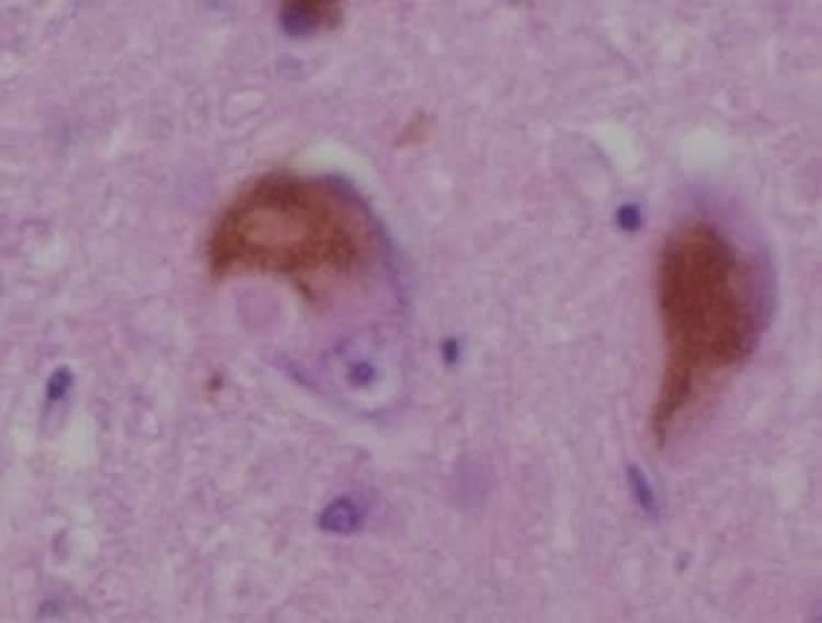
Parkinson’s disease is one of a number of neurodegenerative diseases caused when naturally occurring proteins fold into the wrong shape and stick together with other proteins, eventually forming thin filament-like structures called amyloid fibrils. These amyloid deposits of aggregated alpha-synuclein, also known as Lewy bodies, are the hallmark of Parkinson’s disease.
Parkinson’s disease is the second-most common neurodegenerative disease worldwide (after Alzheimer’s disease). Close to 130,000 people in the UK, and more than seven million worldwide, have the disease. Symptoms include muscle tremors, stiffness and difficulty walking. Dementia is common in later stages of the disease.
“What hasn’t been clear is whether once alpha-synuclein fibrils have formed they are still toxic to the cell,” said Dr Dorothea Pinotsi of Cambridge’s Department of Chemical Engineering and Biotechnology, the paper’s first author.
Pinotsi and her colleagues from Cambridge’s Department of Chemical Engineering & Biotechnology and Department of Chemistry, and led by Dr Gabriele Kaminski Schierle, have used optical ‘super-resolution’ techniques to look into live neurons without damaging the tissue. “Now we can look at how proteins associated with neurodegenerative conditions grow over time, and how these proteins come together and are passed on to neighbouring cells,” said Pinotsi.
The researchers used different forms of alpha-synuclein and observed their behaviour in neurons from rats. They were then able to correlate what they saw with the amount of toxicity that was present.
They found that when they added alpha-synuclein fibrils to the neurons, they interacted with alpha-synuclein protein that was already in the cell, and no toxic effects were present.
“It was believed that amyloid fibrils that attack the healthy protein in the cell would be toxic to the cell,” said Pinotsi. “But when we added a different, soluble form of alpha-synuclein, it didn’t interact with the protein that was already present in the neuron and interestingly this was where we saw toxic effects and cells began to die. So somehow, when the soluble protein was added, it created this toxic effect. The damage appears to be done before visible fibrils are even formed.”
The researchers then observed that by adding the soluble form of alpha-synuclein together with amyloid fibrils, the toxic effect of the former could be overcome. It appeared that the amyloid fibrils acted like magnets for the soluble protein and mopped up the soluble protein pool, shielding against the associated toxic effects.
“These findings change the way we look at the disease, because the damage to the neuron can happen when there is simply extra soluble protein present in the cell – it’s the excess amount of this protein that appears to cause the toxic effects that lead to the death of brain cells,” said Pinotsi. Extra soluble protein can be caused by genetic factors or ageing, although there is some evidence that it could also be caused by trauma to the head.
The research shows how important it is to fully understand the processes at work behind neurodegenerative diseases, so that the right step in the process can be targeted. “With these optical super-resolution techniques, we can really see details we couldn’t see before, so we may be able to counteract this toxic effect at an early stage,” said Pinotsi.
Funding: The research was funded by the Medical Research Council, the Engineering and Physical Sciences Research Council, and the Wellcome Trust.
Source: Sarah Collins – University of Cambridge
Image Source: The image is credited to Dr. Andreas Becker and is licensed CC BY SA 3.0.
Original Research: The study will appear in PNAS during the week of March 14 2016.


