

Montreal Neurological Institute scientists shed light on the mechanism that could be driving the spread of Parkinson’s along neuronal highways.
Scientists at the Montreal Neurological Institute and Hospital -The Neuro, at McGill University and the McGill University Health Centre, have made advances in understanding the process involved in the progression and spread of Parkinson’s disease (PD) within the brain.
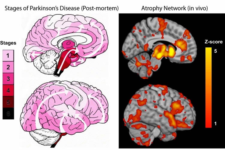
The study, published in September issue of eLife, focused on understanding the process that drives the disease’s progression by mapping the distribution and degree of atrophy, characteristic of the disease, in certain brain regions and identify the paths leading the spread from affected to healthy tissue.
“Past studies have failed to consistently demonstrate regional brain atrophy in earlier stages of the disease due to samples of subjects that were too small and to methods that were less sensitive in detecting all aspects of the disease’s impact on the brain. We now have the means to map the disease with greater sensitivity than previously possible,” says Dr. Alain Dagher, senior author of the study.
The researchers had access to an unprecedented number of MRI scans and clinical data through the open source Parkinson’s Progression Markers Initiative (PPMI) database. Thanks to this wealth of data, researchers were able to analyse MRI scans which show the structure of the brains of 230 people in the early stages of Parkinson’s disease and compare them to those from age-matched healthy individuals. This allowed them to identify the set of brain regions that show atrophy in the early stages of the disease.
“The atrophy pattern on MRI is compatible with a disease process that spreads via brain networks – something that had never been shown in human patients before, and would support the hypothesis that PD is caused by a “toxic agent” that spreads trans-neuronally,” says Dr. Alain Dagher.
The findings add new evidence to the hypothesis that brain cells in Parkinson’s patients might deteriorate according to a prion-like mode of disease propagation, in which a toxic agent spreads from brain cell to brain cell utilizing the normal connections of the brain. Similar mechanisms have been proposed for diseases ranging from Alzheimer’s Disease to Bovine Spongiform Encephalopathy. The process would involve the spread of alpha-synuclein, a toxic misfolded protein with the ability to make copies of itself and infect neighbouring cells while traveling through the brain’s neuronal highways.
The patients who were enrolled in this study will continue to be evaluated on a yearly basis providing researchers with more data to continue mapping how the disease progresses throughout the brain and furthering our understanding of its causes.
Current treatment options help control or minimize symptoms including tremors, slowness of movement, stiffness or rigidity, and loss of balance. The findings of this study hold exciting therapeutic implications. In the longer term, it will help researchers develop new techniques to assess the efficacy of drugs that could target the culprit protein and might eventually lead to treatments that will prevent, slow, halt or even reverse the progression of PD.
PPMI is an observational clinical study to verify progression markers in Parkinson’s disease. PPMI has emerged as a model for following multiple cohorts of significant interest and is being conducted at a network of clinical sites around the world. The study is designed to establish a comprehensive set of clinical, imaging and biosample data that will be used to define biomarkers of PD progression. Once these biomarkers are defined, they can be used in therapeutic studies, which is the ultimate goal.
Funding: The study, conducted by Alain Dagher and collaborators and the Montreal Neurological Institute, was supported by grants from the Michael J Fox Foundation for Parkinson’s Research, the W. Garfield Weston Foundation, and the Alzheimer’s Association, the Canadian Institutes for Health Research, and the Natural Sciences and Engineering Research Council of Canada.
Source: Maya-Olivia Eyssen – McGill University
Image Credit: The image is credited to the researchers/McGill University
Original Research: Abstract for “Network structure of brain atrophy in de novo Parkinson’s Disease” by Yashar Zeighami, Miguel Ulla, Yasser Iturria-Medina, Mahsa Dadar, Yu Zhang, Kevin Michel-Herve Larcher, Vladimir Fonov, Alan C Evans, Douglas Louis Collins, and Alain Dagher in eLife. Published online September 7 2015 doi:10.7554/eLife.08440
Abstract
Network structure of brain atrophy in de novo Parkinson’s Disease
We mapped the distribution of atrophy in Parkinson’s Disease (PD) using MRI and clinical data from 232 PD patients and 117 controls from the Parkinson’s Progression Markers Initiative. Deformation based morphometry and independent component analysis identified PD-specific atrophy in the midbrain, basal ganglia, basal forebrain, medial temporal lobe, and discrete cortical regions. The degree of atrophy reflected clinical measures of disease severity. The spatial pattern of atrophy demonstrated overlap with intrinsic networks present in healthy brain, as derived from functional MRI. Moreover, the degree of atrophy in each brain region reflected its functional and anatomical proximity to a presumed disease epicenter in the substantia nigra, compatible with a trans-neuronal spread of the disease. These results support a network-spread mechanism in PD. Finally, the atrophy pattern in PD was also seen in healthy aging, where it also correlated with the loss of striatal dopaminergic innervation.
“Network structure of brain atrophy in de novo Parkinson’s Disease” by Yashar Zeighami, Miguel Ulla, Yasser Iturria-Medina, Mahsa Dadar, Yu Zhang, Kevin Michel-Herve Larcher, Vladimir Fonov, Alan C Evans, Douglas Louis Collins, and Alain Dagher in eLife. Published online September 7 2015 doi:10.7554/eLife.08440


