Optogenetically stimulating mice’s brains five days after stroke improved the animals’ motor control and brain biochemistry.
When investigators at the Stanford University School of Medicine applied light-driven stimulation to nerve cells in the brains of mice that had suffered strokes several days earlier, the mice showed significantly greater recovery in motor ability than mice that had experienced strokes but whose brains weren’t stimulated.
These findings, published online Aug. 18 in Proceedings of the National Academy of Sciences, could help identify important brain circuits involved in stroke recovery and usher in new clinical therapies for stroke, including the placement of electrical brain-stimulating devices similar to those used for treating Parkinson’s disease, chronic pain and epilepsy. The findings also highlight the neuroscientific strides made possible by a powerful research technique known as optogenetics.
Stroke, with 15 million new victims per year worldwide, is the planet’s second-largest cause of death, according to Gary Steinberg, MD, PhD, professor and chair of neurosurgery and the study’s senior author. In the United States, stroke is the largest single cause of neurologic disability, accounting for about 800,000 new cases each year — more than one per minute — and exacting an annual tab of about $75 billion in medical costs and lost productivity.

The only approved drug for stroke in the United States is an injectable medication called tissue plasminogen activator, or tPA. If infused within a few hours of the stroke, tPA can limit the extent of stroke damage. But no more than 5 percent of patients actually benefit from it, largely because by the time they arrive at a medical center the damage is already done. No pharmacological therapy has been shown to enhance recovery from stroke from that point on.
Enhancing recovery
But in this study — the first to use a light-driven stimulation technology called optogenetics to enhance stroke recovery in mice — the stimulations promoted recovery even when initiated five days after stroke occurred.
“In this study, we found that direct stimulation of a particular set of nerve cells in the brain — nerve cells in the motor cortex — was able to substantially enhance recovery,” said Steinberg, the Bernard and Ronni Lacroute-William Randolph Hearst Professor in Neurosurgery and Neurosciences.
About seven of every eight strokes are ischemic: They occur when a blood clot cuts off oxygen flow to one or another part of the brain, destroying tissue and leaving weakness, paralysis and sensory, cognitive and speech deficits in its wake. While some degree of recovery is possible — this varies greatly among patients depending on many factors, notably age — it’s seldom complete, and typically grinds to a halt by three months after the stroke has occurred.
Animal studies have indicated that electrical stimulation of the brain can improve recovery from stroke. However, “existing brain-stimulation techniques activate all cell types in the stimulation area, which not only makes it difficult to study but can cause unwanted side effects,” said the study’s lead author, Michelle Cheng, PhD, a research associate in Steinberg’s lab.
For the new study, the Stanford investigators deployed optogenetics, a technology pioneered by co-author Karl Deisseroth, MD, PhD, professor of psychiatry and behavioral sciences and of bioengineering. Optogenetics involves expressing a light-sensitive protein in specifically targeted brain cells. Upon exposure to light of the right wavelength, this light-sensitive protein is activated and causes the cell to fire.
Steinberg’s team selectively expressed this protein in the brain’s primary motor cortex, which is involved in regulating motor functions. Nerve cells within this cortical layer send outputs to many other brain regions, including its counterpart in the brain’s opposite hemisphere. Using an optical fiber implanted in that region, the researchers were able to stimulate the primary motor cortex near where the stroke had occurred, and then monitor biochemical changes and blood flow there as well as in other brain areas with which this region was in communication. “We wanted to find out whether activating these nerve cells alone can contribute to recovery,” Steinberg said.
Walking farther
By several behavioral, blood flow and biochemical measures, the answer two weeks later was a strong yes. On one test of motor coordination, balance and muscular strength, the mice had to walk the length of a horizontal beam rotating on its axis, like a rotisserie spit. Stroke-impaired mice whose primary motor cortex was optogenetically stimulated did significantly better in how far they could walk along the beam without falling off and in the speed of their transit, compared with their unstimulated counterparts.
The same treatment, applied to mice that had not suffered a stroke but whose brains had been similarly genetically altered and then stimulated just as stroke-affected mice’s brains were, had no effect on either the distance they travelled along the rotating beam before falling off or how fast they walked. This suggests it was stimulation-induced repair of stroke damage, not the stimulation itself, yielding the improved motor ability.
Stroke-affected mice whose brains were optogenetically stimulated also regained substantially more of their lost weight than unstimulated, stroke-affected mice. Furthermore, stimulated post-stroke mice showed enhanced blood flow in their brain compared with unstimulated post-stroke mice.
In addition, substances called growth factors, produced naturally in the brain, were more abundant in key regions on both sides of the brain in optogenetically stimulated, stroke-affected mice than in their unstimulated counterparts. Likewise, certain brain regions of these optogenetically stimulated, post-stroke mice showed increased levels of proteins associated with heightened ability of nerve cells to alter their structural features in response to experience — for example, practice and learning. (Optogenetic stimulation of the brains of non-stroke mice produced no such effects.)
Steinberg said his lab is following up to determine whether the improvement is sustained in the long term. “We’re also looking to see if optogenetically stimulating other brain regions after a stroke might be equally or more effective,” he said. “The goal is to identify the precise circuits that would be most amenable to interventions in the human brain, post-stroke, so that we can take this approach into clinical trials.”
The study was funded by the National Institute of Neurological Disorders and Stroke (grant 1R21NS082894), Russell and Elizabeth Siegelman, and Bernard and Ronni Lacroute. Other Stanford co-authors of the study are research assistant Eric Wang; research assistant Guohua Sun, MD, PhD; postdoctoral scholar Ahmet Arac, MD; research associate Alex Lee, PhD; medical student Lief Fenno, PhD; and undergraduate students Wyatt Woodson and Stephanie Wang.
Source: Bruce Goldman – Stanford
Contact: Stanford press release
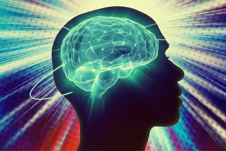
Image Source: The image is credited to Jose-Luis Olivares/MIT and is adapted from a previously published MIT press release on NeuroscienceNews.com
Original Research: Abstract for “Optogenetic neuronal stimulation promotes functional recovery after stroke” by Michelle Y. Cheng, Eric H. Wang, Wyatt J. Woodson, Stephanie Wang, Guohua Sun, Alex G. Lee, Ahmet Arac, Lief E. Fenno, Karl Deisseroth, and Gary K. Steinberg in PNAS. Published online August 18 2014 doi:10.1073/pnas.1404109111


