

Scientists at Seattle Children’s Research Institute have discovered an area of the brain that could control a person’s motivation to exercise and participate in other rewarding activities – potentially leading to improved treatments for depression.
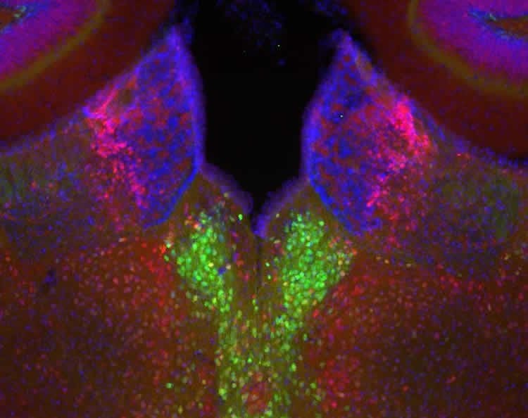
Dr. Eric Turner, a principal investigator in Seattle Children’s Research Institute’s Center for Integrative Brain Research, together with lead author Dr. Yun-Wei (Toni) Hsu, have discovered that a tiny region of the brain – the dorsal medial habenula – controls the desire to exercise in mice. The structure of the habenula is similar in humans and rodents and these basic functions in mood regulation and motivation are likely to be the same across species.
Exercise is one of the most effective non-pharmacological therapies for depression. Determining that such a specific area of the brain may be responsible for motivation to exercise could help researchers develop more targeted, effective treatments for depression.
“Changes in physical activity and the inability to enjoy rewarding or pleasurable experiences are two hallmarks of major depression,” Turner said. “But the brain pathways responsible for exercise motivation have not been well understood. Now, we can seek ways to manipulate activity within this specific area of the brain without impacting the rest of the brain’s activity.”
Dr. Turner’s study, titled “Role of the Dorsal Medial Habenula in the Regulation of Voluntary Activity, Motor Function, Hedonic State, and Primary Reinforcement,” was published today by the Journal of Neuroscience and funded by the National Institute of Mental Health and National Institute on Drug Abuse. The study used mouse models that were genetically engineered to block signals from the dorsal medial habenula. In the first part of the study, Dr. Turner’s team collaborated with Dr. Horacio de la Iglesia, a professor in University of Washington’s Department of Biology, to show that compared to typical mice, who love to run in their exercise wheels, the genetically engineered mice were lethargic and ran far less. Turner’s genetically engineered mice also lost their preference for sweetened drinking water.
“Without a functioning dorsal medial habenula, the mice became couch potatoes,” Turner said. “They were physically capable of running but appeared unmotivated to do it.”
In a second group of mice, Dr. Turner’s team activated the dorsal medial habenula using optogenetics – a precise laser technology developed in collaboration with the Allen Institute for Brain Science. The mice could “choose” to activate this area of the brain by turning one of two response wheels with their paws. The mice strongly preferred turning the wheel that stimulated the dorsal medial habenula, demonstrating that this area of the brain is tied to rewarding behavior.
Past studies have attributed many different functions to the habenula, but technology was not advanced enough to determine roles of the various subsections of this area of the brain, including the dorsal medial habenula.
“Traditional methods of stimulation could not isolate this part of the brain,” Turner said. “But cutting-edge technology at Seattle Children’s Research Institute makes discoveries like this possible.”
As a professor in the University of Washington Department of Psychiatry and Behavioral Sciences, Dr. Turner treats depression and hopes this research will make a difference in the lives of future patients.
“Working in mental health can be frustrating,” Turner said. “We have not made a lot of progress in developing new treatments. I hope the more we can learn about how the brain functions the more we can help people with all kinds of mental illness.”
Contact: Stacey DiNuzzo – Seattle Children’s Hospital
Source: Seattle Children’s Hospital press release
Image Source: The image is credited to the NIH and is in the public domain
Original Research: Abstract for “Role of the Dorsal Medial Habenula in the Regulation of Voluntary Activity, Motor Function, Hedonic State, and Primary Reinforcement” by Yun-Wei A. Hsu, Si D. Wang, Shirong Wang, Glenn Morton, Hatim A. Zariwala, Horacio O. de la Iglesia, and Eric E. Turner in Journal of Neuroscience. Published online August 20 2014 doi:10.1523/JNEUROSCI.1861-14.2014


