

Summary: Researchers have mapped cells that form the brain’s blood vessels and blood brain barrier.
Source: Karolinska Institute.
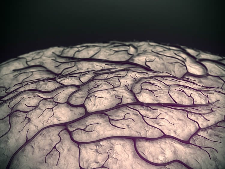
Even though diseases of the brain vasculature are some of the most common causes of death in the West, knowledge of these blood vessels is limited. Now researchers from Uppsala University and Karolinska Institutet present in the journal Nature a detailed molecular atlas of the cells that form the brain’s blood vessels and the life-essential blood-brain barrier. The atlas provides new knowledge regarding the functions of the cells and the barrier, and clues to which cell types are involved in different diseases.
The vasculature of the brain is, like elsewhere on the body, made up of arteries, veins and thin, intervening vessels called capillaries through which the main exchange of oxygen, nutrients and waste products takes place. However, the vessels of the brain differ from others in one important respect – the so-called blood-brain barrier, which acts as a filter that blocks certain substances from passing through the vessel walls, thus protecting the brain from potentially toxic products while letting through whatever it needs for its structure and function.
“It is becoming increasingly clear that a fully functional blood-brain barrier is essential to brain health and that a dysfunctional barrier is a factor of many brain diseases,” says study leader Christer Betsholtz, professor at Uppsala University and Karolinska Institutet’s Department of Medicine, Huddinge. “The structure of the blood-brain barrier hasn’t been fully known, and so a detailed atlas of the brain’s vasculature and its barrier functionality is needed.”
Cellular and molecular atlas
Using a relatively new technique called single cell RNA sequencing, the researchers have produced a cellular and molecular atlas of the mouse brain vasculature. The blood vessels were broken apart in individual cells, which were then, one by one, mapped in accordance with their gene expression patterns and compared. The basic cell type and any gradual specialisations could then be ascertained for each cell. Finally, the molecular map was matched with the corresponding anatomy in tissue analyses using specific markers.
“For the first time we’ve been able to show in detail how the blood-brain barrier differs among the various types of brain blood vessels,” says lead author Michael Vanlandewijck, assistant professor at Uppsala University and Karolinska Institutet and director of the Single Cell Analysis Unit at Karolinska Institutet’s and AstraZeneca’s Integrated Cardio Metabolic Centre (ICMC).
It was long thought that the blood-brain barrier was made up of specialised endothelial cells in the blood vessels; the new study shows, however, that there are probably many other types of cells involved in the maintenance of the blood-brain barrier as well, including cells called pericytes in the capillary walls. The researchers were also able to establish the molecular identity of another cell type in the vascular wall, a kind of connective tissue cell located in a narrow space just outside the brain’s blood vessels.
“This space has been posited to act as the brain’s lymph system, so it’ll now be incredibly interesting to study these cells further using the markers we’ve found,” says Dr Vanlandewijck.
Links disease-causing genes to specific cell types
The atlas means that a number of genes with known or presumed function in different brain diseases can now be associated with specific cell types in the brain’s vasculature.
“We already have results indicating that many more cell types than previously thought are involved in neurovascular diseases such as Alzheimer’s and brain tumours,” says Professor Betsholtz. “We’re now able to study this systematically in different diseases with the same type of analyses as we’ve used here.”
Funding: The study was conducted by researchers at Uppsala University and Karolinska Institutet and colleagues in France, Finland, Switzerland, Japan and China. It was financed by AstraZeneca, the Swedish Research Council, the European Research Council (ERC), the Leducq Foundation, the Swedish Cancer Society, the Knut and Alice Wallenberg Foundation, the Swedish Brain Fund, the Swiss National Science Foundation and the Synapsis Foundation.
Source: Karolinska Institute
Publisher: Organized by NeuroscienceNews.com.
Image Source: NeuroscienceNews.com image is adapted from the Karolinska Institute news release.
Original Research: Abstract in Nature.
doi:10.1038/nature25739
[cbtabs][cbtab title=”MLA”]Karolinska Institute “Atlas of Brain Blood Vessels Provides Fresh Clues to Brain Diseases.” NeuroscienceNews. NeuroscienceNews, 15 February 2018.
<https://neurosciencenews.com/neurology-brain-blood-vessels-8494/>.[/cbtab][cbtab title=”APA”]Karolinska Institute (2018, February 15). Atlas of Brain Blood Vessels Provides Fresh Clues to Brain Diseases. NeuroscienceNews. Retrieved February 15, 2018 from https://neurosciencenews.com/neurology-brain-blood-vessels-8494/[/cbtab][cbtab title=”Chicago”]Karolinska Institute “Atlas of Brain Blood Vessels Provides Fresh Clues to Brain Diseases.” https://neurosciencenews.com/neurology-brain-blood-vessels-8494/ (accessed February 15, 2018).[/cbtab][/cbtabs]
Abstract
A molecular atlas of cell types and zonation in the brain vasculature
Cerebrovascular disease is the third most common cause of death in developed countries, but our understanding of the cells that compose the cerebral vasculature is limited. Here, using vascular single-cell transcriptomics, we provide molecular definitions for the principal types of blood vascular and vessel-associated cells in the adult mouse brain. We uncover the transcriptional basis of the gradual phenotypic change (zonation) along the arteriovenous axis and reveal unexpected cell type differences: a seamless continuum for endothelial cells versus a punctuated continuum for mural cells. We also provide insight into pericyte organotypicity and define a population of perivascular fibroblast-like cells that are present on all vessel types except capillaries. Our work illustrates the power of single-cell transcriptomics to decode the higher organizational principles of a tissue and may provide the initial chapter in a molecular encyclopaedia of the mammalian vasculature.


