UT Arlington researchers have successfully used a portable brain-mapping device to show limited prefrontal cortex activity among student veterans with Post Traumatic Stress Disorder when they were asked to recall information from simple memorization tasks.
The study by bioengineering professor Hanli Liu and Alexa Smith-Osborne, an associate professor of social work, and two other collaborators was published in the May 2014 edition of NeuroImage: Clinical. The team used functional near infrared spectroscopy to map brain activity responses during cognitive activities related to digit learning and memory retrial.
Smith-Osborne has used the findings to guide treatment recommendations for some veterans through her work as principal investigator for UT Arlington’s Student Veteran Project, which offers free services to veterans who are undergraduates or who are considering returning to college.
“When we retest those student veterans after we’ve provided therapy and interventions, they’ve shown marked improvement,” Smith-Osborne said. “The fNIRS data have shown improvement in brain functions and responses after the student veterans have undergone treatment.”
“It also shows how PTSD can affect the way we learn and our ability to recall information, so this new way of brain imaging advances our understanding of PTSD.” Liu said.
This study is multi-disciplinary, associating objective brain imaging with neurological disorders and social work.
While UT Arlington bioengineering faculty associate Fenghua Tian is the primary author assisted by bioengineering graduate research assistant Amarnath Yennu, collaborators of the study include UT Austin psychology professor Francisco Gonzalez-Lima and psychology professor Carol North with UT Southwestern Medical Center and the Veterans Administration North Texas Health Care System.
Khosrow Behbehani, dean of the UT Arlington College of Engineering, said this collaborative research is “allowing the researchers to objectively measure the changes in the level of oxygen in the brain and relate them to some of the brain functions that may have been adversely affected by trauma or stress.”
Numerous neuropsychological studies have linked learning dysfunctions – such as memory loss, attention deficits and learning disabilities – with PTSD.
The new study involved 16 combat veterans previously diagnosed with PTSD who were experiencing distress and functional impairment affecting cognitive and related academic performance. The veterans were directed to perform a series of number-ordering tasks on a computer while researchers monitored their brain activity through near infrared spectroscopy, a noninvasive neuroimaging technology.
The research found that participants with PTSD experienced significant difficulty recalling the given digits compared with a control group. This deficiency is closely associated with dysfunction of a portion in the right frontal cortex. The team also determined that near infrared spectroscopy was an effective tool for measuring cognitive dysfunction associated with PTSD.
With that information, Smith-Osborne said mental healthcare providers could customize a treatment plan best suited for that individual.
“It’s not a one-size-fits-all treatment plan but a concentrated effort to tailor the treatment based on where that person is on the learning scale,” Smith-Osborne said.
Smith-Osborne and Liu hope that their research results lead to better and more comprehensive care for veterans and a better college education.
The work was supported in part by the Hogg Foundation for Mental Health, The National Institutes of Health’s National Institute of Neurological Disorders and Stroke, the UT Arlington Research Enhancement fund, the Department of Education, the Dallas Foundation and private donors in support of the Student Veteran Project.
Contact: Herb Booth – UT Arlington
Source: UT Arlington press release
Image Source: The image is credited to Liu et al/NeuroImage:Clinical and is adapted from the open access research paper, licensed Creative Commons Attribution-NonCommercial-NoDerivs 3.0 Unported
Original Research: Full open access research for “Prefrontal responses to digit span memory phases in patients with post-traumatic stress disorder (PTSD): A functional near infrared spectroscopy study” by Fenghua Tian, Amarnath Yennu, Alexa Smith-Osborne, F. Gonzalez-Lima, Carol S. North, Hanli Liu in NeuroImage: Clinical. Published online June 16 2014 doi:10.1016/j.nicl.2014.05.005
Prefrontal responses to digit span memory phases in patients with post-traumatic stress disorder (PTSD): A functional near infrared spectroscopy study
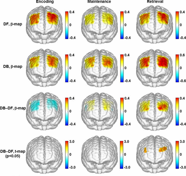
Neuroimaging studies of post-traumatic stress disorder (PTSD)-related memory impairments have consistently implicated abnormal activities in the frontal and parietal lobes. However, most studies have used block designs and could not dissociate the multiple phases of working memory. In this study, the involvement of the prefrontal cortex in working memory phases was assessed among veterans with PTSD and age-/gender-matched healthy controls. Multichannel functional near infrared spectroscopy (fNIRS) was utilized to measure prefrontal cortex hemodynamic activations during memory of neutral (i.e., not trauma-related) forward and backward digit span tasks. An event-related experimental design was utilized to dissociate the different phases (i.e., encoding, maintenance and retrieval) of working memory. The healthy controls showed robust hemodynamic activations during the encoding and retrieval processes. In contrast, the veterans with PTSD were found to have activations during the encoding process, but followed by distinct deactivations during the retrieval process. The PTSD participants, but not the controls, appeared to suppress prefrontal activity during memory retrieval. This deactivation was more pronounced in the right dorsolateral prefrontal cortex during the retrieval phase. These deactivations in PTSD patients might implicate an active inhibition of dorsolateral prefrontal neural activity during retrieval of working memory.
“Prefrontal responses to digit span memory phases in patients with post-traumatic stress disorder (PTSD): A functional near infrared spectroscopy study” by Fenghua Tian, Amarnath Yennu, Alexa Smith-Osborne, F. Gonzalez-Lima, Carol S. North, Hanli Liu in NeuroImage: Clinical, June 16 2014 doi:10.1016/j.nicl.2014.05.005