

Sometimes, a new way of looking at something can bring to light an entirely new perspective.
Using a different type of MRI imaging, researchers at the University of Iowa have discovered previously unrecognized differences in the brains of patients with bipolar disorder. In particular, the study, published Jan. 6 in the journal Molecular Psychiatry, revealed differences in the white matter of patients’ brains and in the cerebellum, an area of the brain not previously linked with the disorder. Interestingly, the cerebellar differences were not present in patients taking lithium, the most commonly used treatment for bipolar disorder.
“This imaging technique appears to be sensitive to things that just have not been imaged effectively before. So it’s really providing a new picture and new insight into the composition and function of the brain [in bipolar disease],” says John Wemmie, MD, PhD, UI professor of psychiatry and senior study author.
Bipolar disorder affects about 1 percent of the population. Despite being relatively common, scientists do not have a good understanding of what causes this psychiatric condition, which is characterized by sudden mood shifts from normal to depressed or to an abnormally elevated or “manic” mood state.
The study examined 15 patients with bipolar disorder and 25 control subjects matched for age and gender. The bipolar patients were all in normal (euthymic) mood state during the study.
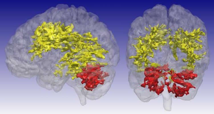
The UI team imaged the participants’ brains using an MRI approach known as quantitative high-resolution T1 rho mapping, which is sensitive to certain byproducts of cell metabolism, including levels of glucose and acidity in the brain. Compared to the brains of people without bipolar disorder, the researchers found that the MRI signal was elevated in the cerebral white matter and the cerebellar region of patients affected by bipolar disorder. The elevated signal may be due to either a reduction in pH or a reduction in glucose concentration – both factors influenced by cell metabolism.
Previous research has suggested that abnormal cell metabolism may play a role in bipolar disorder. However, investigating metabolic abnormalities in the brain has been hindered by lack of a good imaging tools. Available methods are slow, low-resolution, and require researchers to identify the region of interest at the beginning of the study. In contrast, the new imaging approach can rapidly acquire a high-resolution image of the whole brain. The study is the first time this MRI technique has been used to investigate a psychiatric disease.
One reason researchers didn’t know that the cerebellum might be important in bipolar disorder, is because no one chose to look there, says Casey Johnson, PhD, UI postdoctoral researcher and first author on the study.
“Our study was essentially exploratory. We didn’t know what we would find,” he adds. “The majority of bipolar disorder research has found differences in the frontal region of the brain. We found focal differences in the cerebellum, which is a region that hasn’t really been highlighted in the bipolar literature before.”
Spurred on by the finding, Johnson and Wemmie conducted an extensive search of the scientific literature on bipolar disorder and began to find pieces of evidence that suggested that the cerebellum may function abnormally in bipolar disorder and that lithium might potentially target the cerebellum and alter glucose levels in this brain region.
“Our paper, with this new technique, starts to bring all these pieces of evidence together for the first time,” Johnson says.
Wemmie hopes that the new insights provided by the T1 rho imaging might help refine understanding of the abnormalities that underlie bipolar disease and lead to better ways to diagnose and treat this problem.
While lithium can be an effective mood stabilizer for people with bipolar disorder, it causes numerous unpleasant side effects for patients.
“If lithium’s effect on the cerebellum is the key to its effectiveness as a mood stabilizer, then a more targeted treatment that causes the same change in the cerebellum without affecting other systems might be a better treatment for patients with bipolar disorder,” Wemmie says.
In addition to Wemmie and Johnson, the study team included UI researcher Jess Fiedorowicz, Vincent Magnotta, Robin Follmer, Ipek Oguz, Lois Warren, and Gary Christensen.
A major source of funding for the research was a philanthropic gift from UI alumnus Roger Koch, who established the Roger L. Koch Mental Illness Research Fund in 2010 through the University of Iowa Foundation.
The study was also supported by grants from the National Institutes of Health, the Department of Veterans Affairs, and NARSAD.
Contact: Jennifer Brown – University of Iowa Health Care
Source: University of Iowa Health Care press release
Image Source: The image is credited to the researchers/University of Iowa and is adapted from the press release
Original Research: Abstract for “Brain abnormalities in bipolar disorder detected by quantitative T1ρ mapping” by C P Johnson, R L Follmer, I Oguz, L A Warren, G E Christensen, J G Fiedorowicz, V A Magnotta and J A Wemmie in Molecular Psychiatry. Published online January 6 2015 doi:10.1038/mp.2014.157


