The motor neuron disease Amyotrophic lateral sclerosis (ALS), also known as Lou Gehrig’s Disease, progresses in a stepwise, sequential pattern which can be classified into four distinct stages, report pathologists with the Perelman School of Medicine at the University of Pennsylvania in the Annals of Neurology.
This post-mortem staging of ALS brain and spinal cord tissues shows, for the first time, how the fatal degenerative disease may progress from one starting point in the central nervous system to other regions of brain and spinal cord. As evidence mounts showing that other neurodegenerative diseases, such as Alzheimer’s and Parkinson’s, transmit disease-specific pathological proteins from cell-to-cell within the interconnected brain regions of affected individuals, this study suggests that a similar transmission process may be spreading toxic TDP-43 proteins and damaging motor neurons in the brain and spinal cord of ALS patients.
“Researchers at Penn and beyond can now start to investigate and try to confirm this transmission process in ALS, by looking for cell to cell transmission of TDP-43 in models of ALS,” said senior author John Trojanowski, MD, PhD, director of the Penn Institute on Aging and professor of Pathology and Laboratory Medicine at Penn. “If this transmission process can be identified and understood in ALS, this opens a new pathway for treatments, such as immunotherapies that could interrupt or block damaged and destructive proteins from corrupting other cells, thus halting the spread of disease.”

Researchers examined 76 autopsies of patients with ALS, measured pathological TPD-43 distribution and concentration in the nervous system, and were able to map out four distinct stages to assess the disease and burden of pathology.
- In stage 1, TDP-43 pathology was found within the primary motor cortex, as well as neurons in the spinal cord and nerves in the brainstem involved with swallowing, breathing and movement.
- Stage 2 added to stage 1, with TDP-43 progressing forward in the brain and into brainstem areas important for balance and posture.
- In stage 3, TDP-43 pathology moved further forward in the frontal cortex and also to areas just behind the primary motor cortex.
- By stage 4, TDP-43 lesions spread more widely to the temporal lobe and hippocampus, areas involved in memory and language comprehension.
The team evaluated genetic material in all but one of the ALS cases, and found that patients with a C9orf72 mutation had a shorter duration of disease (an average of 24 months vs 34 months in patients without the mutation), and while they had the same distribution pattern as ALS cases without the mutation, there was a greater buildup of pathology in each of the regions. 71 percent of patients with this C9orf72 mutation had a family history of ALS, frontotemporal degeneration (FTD) or another neurodegenerative disease, and 29 percent of cases with the C9orf72 mutation were apparently sporadic cases, lacking any ALS-linked genetic mutation.
The information gathered in these post-mortem autopsies mirrors what neurologists see in patients clinically. Future efforts may interrogate the process of disease progression further, work to identify biomarkers that can help detect the burden of disease in patients during life, and, most importantly, home in on therapies to arrest the progression of ALS.
Notes about this ALS and neurodegeneration research
Lead author of the study, Johannes Brettschneider, MD, is a visiting professor in the Center for Neurodegenerative Disease Research at the University of Pennsylvania, from the department of Neurology at the University of Ulm in Germany. The research team also includes experts from Penn’s Center for Neurodegenerative Disease Research (Jon Toledo, MD, John Robinson, BS, David Irwin, MD, EunRan Suh, PhD, Vivianna Van Deerlin, MD, PhD, Elizabeth Wood, MS, Young Baek, MS, Linda Kwong, PhD, Edward Lee, MD, PhD, and Virginia M-Y Lee, PhD, MBA) and from the department of Neurology (Lauren Elman, MD, and Leo McCluskey, MD), along with colleagues from the University of Ulm.
The study was funded by the National Institutes of Health (AG033101, AG017586, AG010124, AG032953, AG039510, NS044266, AG000255), Wyncote Foundation, Koller Family Foundation, and Deutsche Forschungsgemeinschaft.
Contact: Kim Menard – University of Pennsylvania
Source: University of Pennsylvania press release
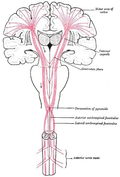
Image Source: The brain and CNS diagram is credited to Gray’s Anatomy and is in the public domain.
Original Research: Abstract for “Stages of pTDP-43 pathology in amyotrophic lateral sclerosis” by Johannes Brettschneider, Kelly Del Tredici, Jon B. Toledo, John L. Robinson, David J. Irwin, Murray Grossman, EunRan Suh, Vivianna M. Van Deerlin, Elisabeth M. Wood, Young Baek, Linda Kwong, Edward B. Lee, Lauren Elman, Leo McCluskey, Lubin Fang, Simone Feldengut, Albert C. Ludolph, Virginia M.-Y. Lee, Heiko Braak and John Q. Trojanowski in Annals of Neurology. Published online June 19 2013 DOI: 10.1002/ana.23937