

Ever wake at night needing a drink of water and then find your way to the kitchen in the dark without stubbing your toe? Researchers at the University of California, San Diego say they have identified a region of the brain that enables you to do that – and generally helps you navigate the world.
Douglas Nitz, associate professor of cognitive science in the UC San Diego Division of Social Sciences, and graduate student Andrew Alexander worked with rats, aka “navigational geniuses,” recording the firing activity of neurons while the animals ran on a zigzag track in different locations, to show that the retrosplenial cortex appears to be critical in putting together all the information necessary for successfully getting from point A to point B. They describe their findings in a paper in the journal Nature Neuroscience.
The world we and other animals navigate is complex and non-linear, quite unlike the way a proverbial crow flies. The authors say our ability to get around its numerous indirect points depends, at minimum, on mapping our position within the environment, knowing routes that take us between locations, and an awareness of the correct actions to initiate at any given time: turn right, turn left, go straight.
Currently, we know that cells encoding these different forms of spatial knowledge are stored in different neural structures. Place cells and grid cells are neurons in the hippocampal circuit that are responsible for mapping the position of an animal with respect to the broader environment. (This is the “inner GPS” system whose discovery, by John O’Keefe, Edvard Moser and May-Britt Moser, was awarded the 2014 Nobel Prize in Physiology or Medicine). Meanwhile, Nitz and other researchers have found neurons in the parietal cortex that generate complex representations of the animal’s position along a route and cells that encode route-related actions.
“But ultimately,” said Nitz, “these different forms of spatial information, generated in distinct structures, must be combined and related to each other in an organized fashion in order for an agent to effectively move through the world. Our study shows that the retrosplenial cortex is an area of the brain that is simultaneously sensitive to mapping interior and exterior spaces and may be a kind of ‘conjunction junction,’ putting together all the necessary information for successful navigation.”
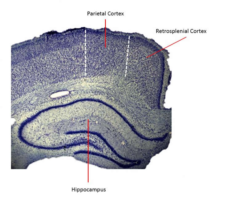
The retrosplenial cortex, which sits in the brain between the parietal and the hippocampus and is vastly interconnected with both regions, has been hypothesized to be critical in this way. The present research, Nitz and Alexander believe, is the first laboratory demonstration of the hypothesis at the level of the cell.
“It’s exciting,” Alexander said, “because it’s the first real evidence of a brain region that’s capable of interfacing really well-known forms of spatial mapping.”
The study’s results are consistent with computational modeling research and with clinical observations of people who have lesions in the retrosplenial, too. Damage to that area can produce problems with episodic memory and create “directional amnesia,” a situation where a person “knows where things are in the world,” Alexander said, “but won’t be able to place the route that they need to get between them.”
The retrosplenial is also one of the very first parts of the brain to degenerate in the early stages of Alzheimer’s, Nitz said. Learning more about the functions of that area of the brain might make it possible for us to devise simple navigation-based tests to spot the disease sooner.
Another potential application of the findings, Nitz said, is in robotics. He is currently collaborating with a colleague at UC Irvine to create an artificial neural network with properties of the retrosplenial cortex so a robot might someday also solve the sorts of navigational problems we (and rats) routinely face.
Funding: The research was supported by the National Science Foundation.
This article was submitted directly to NeuroscienceNews.com by Inga Kiderra at UCSD. We would like to thank Inga for this submission.
Source: Inga Kiderra – UCSD
Image Credit: Image is credited to Andrew Alexander and Douglas Nitz, UC San Diego
Original Research: Abstract for “Retrosplenial cortex maps the conjunction of internal and external spaces” by Andrew S Alexander and Douglas A Nitz in Nature Neuroscience. Published online July 6 2015 doi:10.1038/nn.4058
Abstract
Retrosplenial cortex maps the conjunction of internal and external spaces
Intelligent behavior demands not only multiple forms of spatial representation, but also coordination among the brain regions mediating those representations. Retrosplenial cortex is densely interconnected with the majority of cortical and subcortical brain structures that register an animal’s position in multiple internal and external spatial frames of reference. This unique anatomy suggests that it functions to integrate distinct forms of spatial information and provides an interface for transformations between them. Evidence for this was found in rats traversing two different routes placed at different environmental locations. Retrosplenial ensembles robustly encoded conjunctions of progress through the current route, position in the larger environment and the left versus right turning behavior of the animal. Thus, the retrosplenial cortex has the requisite dynamics to serve as an intermediary between brain regions generating different forms of spatial mapping, a result that is consistent with navigational and episodic memory impairments following damage to this region in humans.
“Retrosplenial cortex maps the conjunction of internal and external spaces” by Andrew S Alexander and Douglas A Nitz in Nature Neuroscience. Published online July 6 2015 doi:10.1038/nn.4058


