

Summary: Researchers identify a new cellular mechanism that may be responsible for multiple sclerosis. Findings could open up new avenues for the development of treatments.
Source: University of Alberta.
A host of possibilities just opened up for treating the mysterious, pervasive illness.
In the relentless battle against multiple sclerosis (MS), U of A researchers recently discovered an entirely new cellular mechanism–an underlying defect in brain cells–that may to be blame for the disease, and a potential hallmark that may be a target for future treatment.
The finding opens the door to a brand new avenue of study in the battle against the cryptic autoimmune disorder that strikes more Canadians than any other nationality worldwide, said Fabrizio Giuliani, study co-author, U of A neurologist and medical director of the Northern Alberta MS Clinic.
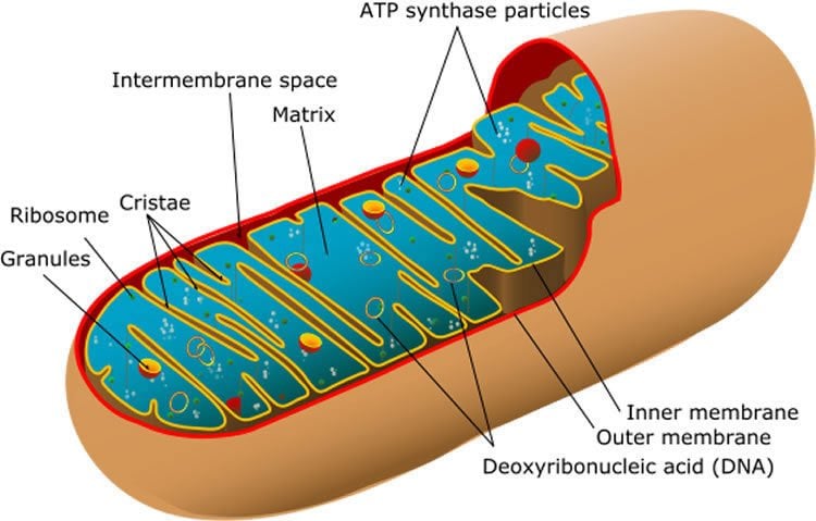
“Scientists have been pointing to the mitochondria, the powerhouse of the cell, as a possible link to MS, but have not been able to decipher how they malfunction. Ours is the first study that combines clinical and lab experiments to explain how mitochondria become defective in MS patients,” said Thomas Simmen, study co-author and cell biology professor.
Specifically, using human brain tissue samples, the researchers discovered how two sub-components within a cell are miscommunicating in patients with MS, and identified how at least one protein (Rab32) is swooping in to trigger the dangerous dysfunction.
“A part of the cell that stores calcium (ER or endoplasmic reticulum) gets too close to the part of the cell that creates energy (mitochondria) when massive amounts of Rab32 are present in the brain of MS patients. The resulting miscommunication with the calcium supply triggers the mitochondria to misbehave, ultimately causing toxicity for brain cells in MS patients,” explained Simmen.
In healthy brain tissue samples, there’s virtually no Rab32 present, he added.
Researchers don’t know why or what causes an unwelcome influx of Rab32 but they theorize the defect could originate at the base of the ER.
With this finding in hand, not only can scientists search for effective treatments that target Rab32, added Simmen, they can embark on determining whether there are other proteins that may be at play.
“Rab32 is just one of the proteins that is having the effect of drawing the ER and mitochondria too close. There are dozens of other possibilities,” he said.
This discovery may give hope to the 100,000 Canadians living with MS who have had to rely on partially effective treatments that aim to reduce inflammation, and, sometimes, controversial experiential treatments such as the vein-widening treatment.
The study, conducted with researchers at the University of Exeter, was recently published in the Journal of Neuroinflammation.
Funding: The Canadian Institutes of Health Research supported the research.
Source: Ross Neitz – University of Alberta
Image Source: NeuroscienceNews.com image is in the public domain.
Original Research: Full open access research for “Rab32 connects ER stress to mitochondrial defects in multiple sclerosis” by Yohannes Haile, Xiaodan Deng, Carolina Ortiz-Sandoval, Nasser Tahbaz, Aleksandra Janowicz, Jian-Qiang Lu, Bradley J. Kerr, Nicholas J. Gutowski, Janet E. Holley, Paul Eggleton, Fabrizio Giuliani and Thomas Simmen is in Journal of Neuroinflammation. Published online January 23 2017 doi:10.1186/s12974-016-0788-z
[cbtabs][cbtab title=”MLA”]University of Alberta “Possible New Treatment for Multiple Sclerosis.” NeuroscienceNews. NeuroscienceNews, 23 March 2017.
<https://neurosciencenews.com/mitochondria-multiple-sclerosis-6279/>.[/cbtab][cbtab title=”APA”]University of Alberta (2017, March 23). Possible New Treatment for Multiple Sclerosis. NeuroscienceNew. Retrieved March 23, 2017 from https://neurosciencenews.com/mitochondria-multiple-sclerosis-6279/[/cbtab][cbtab title=”Chicago”]University of Alberta “Possible New Treatment for Multiple Sclerosis.” https://neurosciencenews.com/mitochondria-multiple-sclerosis-6279/ (accessed March 23, 2017).[/cbtab][/cbtabs]
Abstract
Rab32 connects ER stress to mitochondrial defects in multiple sclerosis
Background
Endoplasmic reticulum (ER) stress is a hallmark of neurodegenerative diseases such as multiple sclerosis (MS). However, this physiological mechanism has multiple manifestations that range from impaired clearance of unfolded proteins to altered mitochondrial dynamics and apoptosis. While connections between the triggering of the unfolded protein response (UPR) and downstream mitochondrial dysfunction are poorly understood, the membranous contacts between the ER and mitochondria, called the mitochondria-associated membrane (MAM), could provide a functional link between these two mechanisms. Therefore, we investigated whether the guanosine triphosphatase (GTPase) Rab32, a known regulator of the MAM, mitochondrial dynamics, and apoptosis, could be associated with ER stress as well as mitochondrial dysfunction.
Methods
We assessed Rab32 expression in MS patient and experimental autoimmune encephalomyelitis (EAE) tissue, via observation of mitochondria in primary neurons and via monitoring of survival of neuronal cells upon increased Rab32 expression.
Results
We found that the induction of Rab32 and other MAM proteins correlates with ER stress proteins in MS brain, as well as in EAE, and occurs in multiple central nervous system (CNS) cell types. We identify Rab32, known to increase in response to acute brain inflammation, as a novel unfolded protein response (UPR) target. High Rab32 expression shortens neurite length, alters mitochondria morphology, and accelerates apoptosis/necroptosis of human primary neurons and cell lines.
Conclusions
ER stress is strongly associated with Rab32 upregulation in the progression of MS, leading to mitochondrial dysfunction and neuronal death.
“Rab32 connects ER stress to mitochondrial defects in multiple sclerosis” by Yohannes Haile, Xiaodan Deng, Carolina Ortiz-Sandoval, Nasser Tahbaz, Aleksandra Janowicz, Jian-Qiang Lu, Bradley J. Kerr, Nicholas J. Gutowski, Janet E. Holley, Paul Eggleton, Fabrizio Giuliani and Thomas Simmen is in Journal of Neuroinflammation. Published online January 23 2017 doi:10.1186/s12974-016-0788-z


