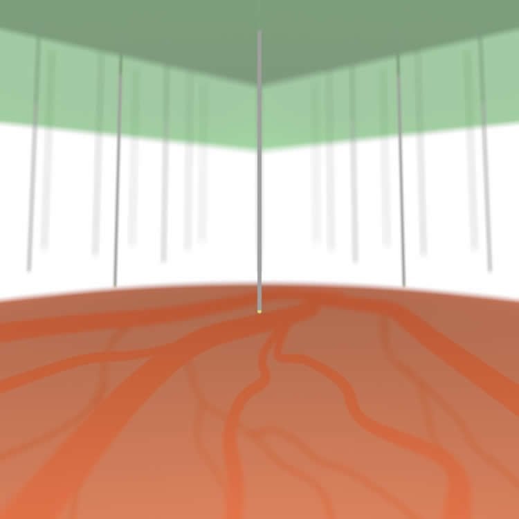
Dissolvable material expands opportunities for flexible microneedles used for brain penetrations.
Microscale needle-electrode array technology has enhanced brain science and engineering applications, such as electrophysiological studies, drug and chemical delivery systems, and optogenetics.
However, one challenge is reducing the tissue/neuron damage associated with needle penetration, particularly for chronic insert experiment and future medical applications. A solution strategy is to use microscale-diameter needles (e.g., < 5 μm) with flexible properties. However, such physically limited needles cannot penetrate the brain and other biological tissues because of needle buckling or fracturing on penetration.

A research team in the Department of Electrical and Electronic Information Engineering and the Electronics-Inspired Interdisciplinary Research Institute (EIIRIS) at Toyohashi University of Technology has developed a methodology to temporarily enhance the stiffness of a long, high-aspect-ratio flexible microneedle (e.g., < 5 μm in diameter and > 500 μm in length), without affecting the needle diameter and flexibility in tissue. This has been accomplished by embedding a needle base in a film scaffold, which dissolves upon contact with biological tissue. Silk fibroin is used as the dissolvable film because it has high biocompatibility, and is a known biomaterial used in implantable devices.
“We investigated preparation of a silk base scaffold for a microneedle, quantitatively analyzed needle stiffness, and evaluated the penetration capability by using mouse brains in vitro/in vivo. In addition, as an actual needle application, we demonstrated fluorescenctce particle depth injection into the brain in vivo,and confirm that by observing fluorescenctce confocal microscope” explained the first author, master’s degree student Satoshi Yagi, and co-author PhD candidate Shota Yamagiwa.
The leader of the research team, Associate Professor Takeshi Kawano said: “Preparation of the dissolvable base scaffold is very simple, but this methodology promises powerful tissue penetrations using numerous high-aspect-ratio flexible microneedles, including recording/stimulation electrodes, glass pipettes, and optogenetic fibers.” He added: “This has the potential to reduce invasiveness drastically and provide safer tissue penetration than conventional approaches.”
Source: Michiteru Kitazaki – Toyohashi University of Technology
Image Credit: The image is credited to Toyohashi University of Technology
Original Research: Abstract for “Dissolvable Base Scaffolds Allow Tissue Penetration of High-Aspect-Ratio Flexible Microneedles” by Satoshi Yagi, Shota Yamagiwa, Yoshihiro Kubota, Hirohito Sawahata, Rika Numano, Tatsuya Imashioya, Hideo Oi, Makoto Ishida, and Takeshi Kawano in Advanced Healthcare Materials. Published online August 6 2015 doi:10.1002/adhm.201500305
Abstract
Dissolvable Base Scaffolds Allow Tissue Penetration of High-Aspect-Ratio Flexible Microneedles
Microscale needle technology is important in electrophysiological studies, drug/chemical delivery systems, optogenetic applications, and so on. In this study, dissolvable needle-base scaffold realizes penetration of high-aspect-ratio flexible microneedles (e.g., 500 μm length) into biological tissues. This methodology, which is applicable to numerous high-aspect-ratio flexible microneedles, should reduce the invasiveness and provide safer tissue penetrations than conventional approaches.
“Dissolvable Base Scaffolds Allow Tissue Penetration of High-Aspect-Ratio Flexible Microneedles” by Satoshi Yagi, Shota Yamagiwa, Yoshihiro Kubota, Hirohito Sawahata, Rika Numano, Tatsuya Imashioya, Hideo Oi, Makoto Ishida, and Takeshi Kawano in Advanced Healthcare Materials. Published online August 6 2015 doi:10.1002/adhm.201500305


