Summary: Researchers have identified a novel signaling system that controls neuroplasticity.
Source: Max Planck Institute.
Researchers from Max Planck Florida Institute for Neuroscience, Duke University, and collaborators have identified a novel signaling system controlling neuronal plasticity.
One of the most fascinating properties of the mammalian brain is its capacity to change throughout life. Experiences, whether studying for a test or experiencing a traumatic situation, alter our brains by modifying the activity and organization of specific neural circuitry, thereby modifying subsequent feelings, thoughts, and behavior. These changes take place in and among synapses, communication junctions between neurons. This experience-driven alteration of brain structure and function is called synaptic plasticity and it is considered the cellular basis for learning and memory.
Many research groups across the globe are dedicated to advancing our understanding of the fundamental principles of learning and memory formation. This understanding is dependent upon identifying the molecules involved in learning and memory and the roles they play in the process. Hundreds of molecules appear to be involved in the regulation of synaptic plasticity, and understanding the interactions among these molecules is crucial to fully understand how memory works.
There are several underlying mechanisms that work together to achieve synaptic plasticity, including changes in the amount of chemical signals released into a synapse and changes in how sensitive a cell’s response is to those signals. In particular, the protein BDNF, its receptor TrkB, and GTPase proteins are involved in some forms of synaptic plasticity, however, very little is known regarding when and where they are activated in the process.
By using sophisticated imaging techniques to monitor the spatiotemporal activation patterns of these molecules in single dendritic spines, the research group led by Dr. Ryohei Yasuda at Max Planck Florida Institute for Neuroscience and Dr. James McNamara at Duke University Medical Center have uncovered critical details of the interplay of these molecules during synaptic plasticity. These exciting findings were published online ahead of print in September 2016 as two independent publications in Nature.

A surprising signaling system within the spine
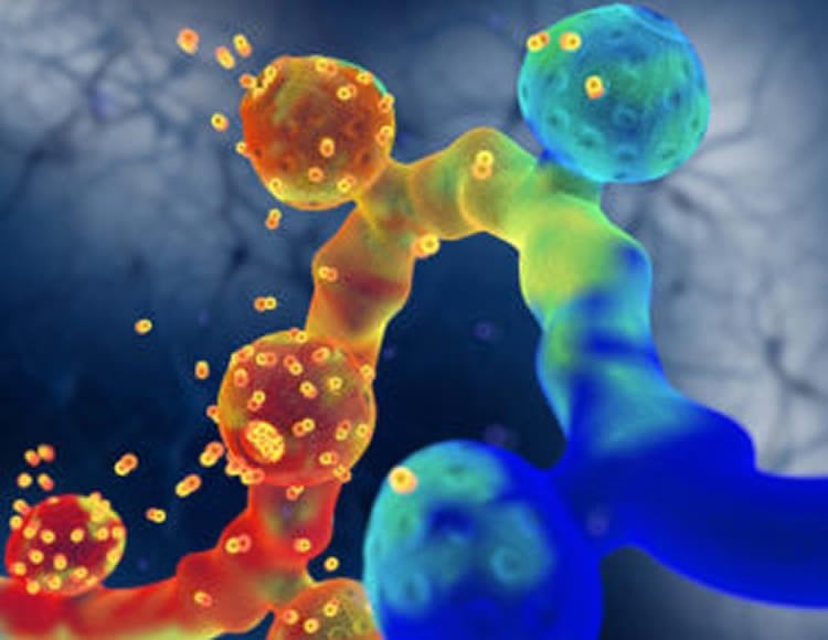
In one of the publications (Harward and Hedrick et al.), the authors identified an autocrine signaling system – a system where molecules act on the same cells that produce them – within single dendritic spines. This autocrine signaling system is achieved by rapid release of the protein, BDNF, from a stimulated spine and subsequent activation of its receptor, TrkB, on the same spine, which further activates signaling inside the spine. This in turn leads to spine enlargement, the process essential for synaptic plasticity. In other words, signaling initiated inside the spine goes outside the spine and activates a receptor on the external surface of the spine, thereby evoking additional signals inside the spine. This finding of an autocrine signaling process within the dendritic spines surprised the scientists.
A spine-autonomous BDNF-TrkB signalling loop critical for synaptic plasticity
What are the consequences of the autocrine signaling within the spine?
The second publication (Hedrick and Harward et al.) reports that the autocrine signaling leads to activation of an additional set of signaling molecules called small GTPase proteins. The findings reveal a three-molecule model of structural plasticity, which implicates the localized, coincident activation of three GTPase proteins Rac1, Cdc42, and RhoA, as a causal feature of structural plasticity. It is known that these proteins regulate the shape of dendritic spines, however, how they work together to control spine structure has remained unclear. The researchers monitored the spatiotemporal activation patterns of these molecules in single dendritic spines during synaptic plasticity and found that all three proteins are activated simultaneously, but their activation patterns differed significantly. One of the differences is that RhoA and Rac1, when activated, spread beyond the stimulated spine to the surrounding dendrite, which facilitates plasticity of surrounding spines. Another difference is that Cdc42 activity was restricted to the stimulated spine, what seems to be necessary to produce spine-specific plasticity. Furthermore, the autocrine BDNF signaling is required for activation of Cdc42 and Rac1, but not for RhoA.
Unprecedented insights into the regulation of synaptic plasticity
These two studies provide unprecedented insights into the regulation of synaptic plasticity. One study revealed for the first time an autocrine signaling system and the second study presented a unique form of biochemical computation in dendrites involving the controlled complementation of three molecules. According to Dr. Yasuda, understanding the molecular mechanisms that are responsible for the regulation of synaptic strength is critical for understanding how neural circuits function, how they form, and how they are shaped by experience. Dr. McNamara noted that disorder of these signaling systems likely underlies dysfunction of synapses that cause epilepsy and a diversity of other diseases of the brain. Because hundreds of species of proteins are involved in the signal transduction that regulates synaptic plasticity, it is essential to investigate the dynamics of more proteins to better understand the signaling mechanisms in dendritic spines.
RhoGTPases: A three-way approach to controlling neural plasticity.
Future research in the Yasuda and McNamara Labs is expected to lead to significant advances in the understanding of intracellular signaling in neurons and will provide key insights into the mechanisms underlying synaptic plasticity and memory formation and brain diseases. These insights will hopefully lead to the development of drugs that could enhance memory and prevent or more effectively treat epilepsy and other brain disorders.
Funding: The research was supported by the National Institutes of Health (F31NS078847, R01NS068410, DP1NS096787, R01NS05621, R01MH080047, R01DA08259, R01HL098351, P01HL096571, and RO1NS030687) the Wakeman Fellowship, and Human Frontier Science Program.
Source: Max Planck Institute
Image Source: NeuroscienceNews.com image is adapted from the MPI press release.
Video Source: The videos are credited to Max Planck Florida Institute for Neuroscience.
Original Research: Abstract for “Rho GTPase complementation underlies BDNF-dependent homo- and heterosynaptic plasticity” by Nathan G. Hedrick, Stephen C. Harward, Charles E. Hall, Hideji Murakoshi, James O. McNamara & Ryohei Yasuda in Nature. Published online September 28 2016 doi:10.1038/nature19784
Abstract for “Autocrine BDNF–TrkB signalling within a single dendritic spine” by Stephen C. Harward, Nathan G. Hedrick, Charles E. Hall, Paula Parra-Bueno, Teresa A. Milner, Enhui Pan, Tal Laviv, Barbara L. Hempstead, Ryohei Yasuda & James O. McNamara in Nature. Published online September 28 2016 doi:10.1038/nature19784
[cbtabs][cbtab title=”MLA”]Max Planck Institute. “Two New Studies Uncover Key Players Responsible for Learning and Memory Formation.” NeuroscienceNews. NeuroscienceNews, 3 October 2016.
<https://neurosciencenews.com/memory-learning-neural-signaling-5193/>.[/cbtab][cbtab title=”APA”]Max Planck Institute. (2016, October 3). Two New Studies Uncover Key Players Responsible for Learning and Memory Formation. NeuroscienceNews. Retrieved October 3, 2016 from https://neurosciencenews.com/memory-learning-neural-signaling-5193/[/cbtab][cbtab title=”Chicago”]Max Planck Institute. “Two New Studies Uncover Key Players Responsible for Learning and Memory Formation.” https://neurosciencenews.com/memory-learning-neural-signaling-5193/ (accessed October 3, 2016).[/cbtab][/cbtabs]
Abstract
Rho GTPase complementation underlies BDNF-dependent homo- and heterosynaptic plasticity
The Rho GTPase proteins Rac1, RhoA and Cdc42 have a central role in regulating the actin cytoskeleton in dendritic spines, thereby exerting control over the structural and functional plasticity of spines and, ultimately, learning and memory. Although previous work has shown that precise spatiotemporal coordination of these GTPases is crucial for some forms of cell morphogenesis, the nature of such coordination during structural spine plasticity is unclear. Here we describe a three-molecule model of structural long-term potentiation (sLTP) of murine dendritic spines, implicating the localized, coincident activation of Rac1, RhoA and Cdc42 as a causal signal of sLTP. This model posits that complete tripartite signal overlap in spines confers sLTP, but that partial overlap primes spines for structural plasticity. By monitoring the spatiotemporal activation patterns of these GTPases during sLTP, we find that such spatiotemporal signal complementation simultaneously explains three integral features of plasticity: the facilitation of plasticity by brain-derived neurotrophic factor (BDNF), the postsynaptic source of which activates Cdc42 and Rac1, but not RhoA; heterosynaptic facilitation of sLTP, which is conveyed by diffusive Rac1 and RhoA activity; and input specificity, which is afforded by spine-restricted Cdc42 activity. Thus, we present a form of biochemical computation in dendrites involving the controlled complementation of three molecules that simultaneously ensures signal specificity and primes the system for plasticity.
“Rho GTPase complementation underlies BDNF-dependent homo- and heterosynaptic plasticity” by Nathan G. Hedrick, Stephen C. Harward, Charles E. Hall, Hideji Murakoshi, James O. McNamara & Ryohei Yasuda in Nature. Published online September 28 2016 doi:10.1038/nature19784
Abstract
Autocrine BDNF–TrkB signalling within a single dendritic spine
Brain-derived neurotrophic factor (BDNF) and its receptor TrkB are crucial for many forms of neuronal plasticity including structural long-term potentiation (sLTP) which is a correlate of an animal’s learning. However, it is unknown whether BDNF release and TrkB activation occur during sLTP, and if so, when and where. Here, using a fluorescence resonance energy transfer-based sensor for TrkB and two-photon fluorescence lifetime imaging microscopy, we monitor TrkB activity in single dendritic spines of CA1 pyramidal neurons in cultured murine hippocampal slices. In response to sLTP induction we find fast (onset < 1 min) and sustained (>20 min) activation of TrkB in the stimulated spine that depends on NMDAR (N-methyl-d-aspartate receptor) and CaMKII signalling and on postsynaptically synthesized BDNF. We confirm the presence of postsynaptic BDNF using electron microscopy to localize endogenous BDNF to dendrites and spines of hippocampal CA1 pyramidal neurons. Consistent with these findings, we also show rapid, glutamate-uncaging-evoked, time-locked BDNF release from single dendritic spines using BDNF fused to superecliptic pHluorin. We demonstrate that this postsynaptic BDNF–TrkB signalling pathway is necessary for both structural and functional LTP. Together, these findings reveal a spine-autonomous, autocrine signalling mechanism involving NMDAR–CaMKII-dependent BDNF release from stimulated dendritic spines and subsequent TrkB activation on these same spines that is crucial for structural and functional plasticity.
“Autocrine BDNF–TrkB signalling within a single dendritic spine” by Stephen C. Harward, Nathan G. Hedrick, Charles E. Hall, Paula Parra-Bueno, Teresa A. Milner, Enhui Pan, Tal Laviv, Barbara L. Hempstead, Ryohei Yasuda & James O. McNamara in Nature. Published online September 28 2016 doi:10.1038/nature19784


