

Luminopsins join glowing + light-sensitive modules.
Optogenetics has taken neuroscience by storm in recent years because the technique allows scientists to study the brain conveniently in animals, activating or inhibiting selected groups of neurons at the flip of a switch. Most often, scientists use a fiber optic cable to deliver light into the brain.
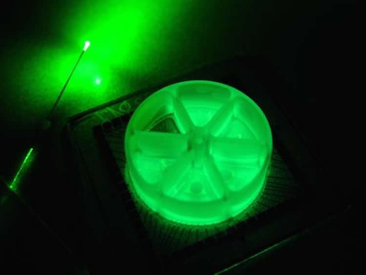
Researchers at Emory and Georgia Tech have developed tools that could allow neuroscientists to put aside the fiber optic cable, and use a glowing protein from coral as the light source instead.
Biomedical engineering student Jack Tung and neurosurgeon/neuroscientist Robert Gross, MD, PhD have dubbed these tools “inhibitory luminopsins” because they inhibit neuronal activity both in response to light and to a chemical supplied from outside.
A demonstration of the luminopsins’ capabilities was published September 24 in the journal Scientific Reports. The authors show that these tools enabled them to modulate neuronal firing, both in culture and in vivo, and modify the behavior of live animals.
Tung and Gross are now using inhibitory luminopsins to study ways to halt or prevent seizure activity in animals.
“We think that this approach may be particularly useful for modeling treatments for generalized seizures and seizures that involve multiple areas of the brain,” Tung says. “We’re also working on making luminopsins responsive to seizure activity: turning on the light only when it is needed, in a closed-loop feedback controlled fashion.”
Gross is a professor of neurosurgery and neurology at Emory University School of Medicine and in the Wallace H. Coulter Department of Biomedical Engineering at Georgia Tech and Emory University. Tung is a MD, PhD student in biomedical engineering. Co-author Claire-Anne Gutekunst, PhD is assistant professor of neurosurgery.
In conventional optogenetics, scientists use genetic engineering techniques to make neurons in animals produce light-sensitive proteins called opsins. These opsins, which come from various algae and salt-tolerant bacteria, respond to a particular wavelength of light by moving ions across the cell membrane – thus stimulating or silencing the neurons where they are expressed. However, fiber optic cables, the usual mode of delivering the light needed to activate the opsins, have a limited reach into the brain, pose a risk of infection and can limit animals’ movements.
To supply light locally and internally, the team took the enzyme luciferase from the soft coral Renilla, which glows in presence of its substrate luciferin, and fused it to an inhibitory opsin – creating a novel fusion protein termed an inhibitory luminopsin (from “luminescent opsin”). An excitatory luminopsin has been reported by other scientists, but its properties were demonstrated only in cultured cells.
To show that luminopsins could modulate behavior in live animals, they injected a gene vector encoding their inhibitory luminopsin into the globus pallidus of rats, on just one side of the brain. Two weeks later, the rats were injected with luciferin. The globus pallidus is involved in motor control, and the presence of luciferin had the effect of disabling the globus pallidus. In response to the drug amphetamine, the animals rotated preferentially in one direction, which mimicked the behavior that results when the globus pallidus is damaged on one side.
“We chose to do the rotation tests because they were simple and easy to measure,” Tung says. “We also wanted to show that expression of luminopsin could be scaled up to multiple structures in the brain.”
The researchers also showed that luminopsins and luciferase together could suppress neural activity in the hippocampus, another region of the brain, in anesthetized rats.
The authors note that similar techniques have been used to stimulate selected neurons in animals with designer drugs, but conclude that luminopsins could be cleaner and easier to use, while offering scientists the choice of influencing neuronal activity with either light or an externally supplied chemical that is otherwise inert.
Funding: This work was funded by NIH/National Institute of Neurological Disorders and Stroke.
Source: Quinn Eastman – Emory Health Sciences
Image Source: The image is credited to Jack Tung
Original Research: Full open access research “Inhibitory luminopsins: genetically-encoded bioluminescent opsins for versatile, scalable, and hardware-independent optogenetic inhibition” by Jack K. Tung, Claire-Anne Gutekunst and Robert E. Gross in Scientific Reports. Published online 25 2015 doi:10.1038/srep14366
Abstract
Inhibitory luminopsins: genetically-encoded bioluminescent opsins for versatile, scalable, and hardware-independent optogenetic inhibition
Optogenetic techniques provide an unprecedented ability to precisely manipulate neural activity in the context of complex neural circuitry. Although the toolbox of optogenetic probes continues to expand at a rapid pace with more efficient and responsive reagents, hardware-based light delivery is still a major hurdle that limits its practical use in vivo. We have bypassed the challenges of external light delivery by directly coupling a bioluminescent light source (a genetically encoded luciferase) to an inhibitory opsin, which we term an inhibitory luminopsin (iLMO). iLMO was shown to suppress action potential firing and synchronous bursting activity in vitro in response to both external light and luciferase substrate. iLMO was further shown to suppress single-unit firing rate and local field potentials in the hippocampus of anesthetized rats. Finally, expression of iLMO was scaled up to multiple structures of the basal ganglia to modulate rotational behavior of freely moving animals in a hardware-independent fashion. This novel class of optogenetic probes demonstrates how non-invasive inhibition of neural activity can be achieved, which adds to the versatility, scalability, and practicality of optogenetic applications in freely behaving animals.
“Inhibitory luminopsins: genetically-encoded bioluminescent opsins for versatile, scalable, and hardware-independent optogenetic inhibition” by Jack K. Tung, Claire-Anne Gutekunst and Robert E. Gross in Scientific Reports. Published online 25 2015 doi:10.1038/srep14366


