

An estimated 8% of Americans will suffer from post traumatic stress disorder (PTSD) at some point during their lifetime. Brought on by an overwhelming or stressful event or events, PTSD is the result of altered chemistry and physiology of the brain. Understanding how threat is processed in a normal brain versus one altered by PTSD is essential to developing effective interventions.
New research from the Center for BrainHealth at The University of Texas at Dallas published online today in Brain and Cognition illustrates how fear arises in the brain when individuals are exposed to threatening images. This novel study is the first to separate emotion from threat by controlling for the dimension of arousal, the emotional reaction provoked, whether positive or negative, in response to stimuli. Building on previous animal and human research, the study identifies an electrophysiological marker for threat in the brain.
“We are trying to find where thought exists in the mind,” explained John Hart, Jr., M.D., Medical Science Director at the Center for BrainHealth. “We know that groups of neurons firing on and off create a frequency and pattern that tell other areas of the brain what to do. By identifying these rhythms, we can correlate them with a cognitive unit such as fear.”
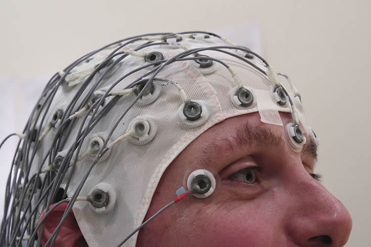
Utilizing electroencephalography (EEG), Dr. Hart’s research team identified theta and beta wave activity that signifies the brain’s reaction to visually threatening images.
“We have known for a long time that the brain prioritizes threatening information over other cognitive processes,” explained Bambi DeLaRosa, study lead author. “These findings show us how this happens. Theta wave activity starts in the back of the brain, in it’s fear center – the amygdala – and then interacts with brain’s memory center – the hippocampus – before traveling to the frontal lobe where thought processing areas are engaged. At the same time, beta wave activity indicates that the motor cortex is revving up in case the feet need to move to avoid the perceived threat.”
For the study, 26 adults (19 female, 7 male), ages 19-30 were shown 224 randomized images that were either unidentifiably scrambled or real pictures. Real pictures were separated into two categories: threatening (weapons, combat, nature or animals) and non-threatening (pleasant situations, food, nature or animals).
While wearing an EEG cap, participants were asked to push a button with their right index finger for real items and another button with their right middle finger for nonreal/scrambled items. Shorter response times were recorded for scrambled images than the real images. There was no difference in reaction time for threatening versus non-threatening images.
EEG results revealed that threatening images evoked an early increase in theta activity in the occipital lobe (the area in the brain where visual information is processed), followed by a later increase in theta power in the frontal lobe (where higher mental functions such as thinking, decision-making, and planning occur). A left lateralized desynchronization of the beta band, the wave pattern associated with motor behavior (like the impulse to run), also consistently appeared in the threatening condition.
This study will serve as a foundation for future work that will explore normal versus abnormal fear associated with an object in other atypical populations including individuals with PTSD.
This work was supported by the Berman Laboratory of Learning and Memory at The University of Texas at Dallas and the Jane and Bud Smith Distinguished Chair.
Contact: Shelly Kirkland – Center for BrainHealth
Source: Center for BrainHealth press release
Image Source: The image is credited to Chris Hope and is licensed Creative Commons Attribution 2.0 Generic
Original Research: Abstract for “Electrophysiological spatiotemporal dynamics during implicit visual threat processing” by Bambi L. DeLaRosa, Jeffrey S. Spence, Scott K.M. Shakal, Michael A. Motes, Clifford S. Calley, Virginia I. Calley, John Hart Jr., and Michael A. Kraut in Brain and Cognition. Published online September 15 2014 doi:10.1016/j.bandc.2014.08.003


