

Summary: Around 15 percent of Ebola survivors have retinal scars that appear to be specific to the disease, a new study reports.
Source: University of Liverpool.
Researchers from the University of Liverpool have conducted a study of Ebola survivors to determine if the virus has any specific effects on the back on the eye using an ultra widefield retinal camera.
To find out more about the broad-ranging symptoms of Post Ebola Syndrome (PES), a clinical research team led by Dr Janet Scott and Dr Calum Semple, from the University’s Institute of Translational Medicine, assessed survivors discharged from the Ebola Treatment Unit at the 34th Regiment Military Hospital in Freetown, Sierra Leone.
Two years on from the Ebola outbreak in West Africa, and many Ebola survivors are still presenting with symptoms of post-Ebola syndrome (PES), including joint and muscle pains and psychiatric and neurological problems.
Hiding viruses
Viruses, like Ebola, can stay hidden in our bodies by exploiting a vulnerability in our immune systems. This vulnerability is called “immune privilege,” and comes from an old observation that foreign tissue transplanted into certain parts of the body don’t elicit the usual immune response. This includes the brain, spinal cord, and eyes. Scientists believe this is because the brain, spinal cord, and eyes are simply too delicate and important to withstand the inflammation that’s typical of an immune response.
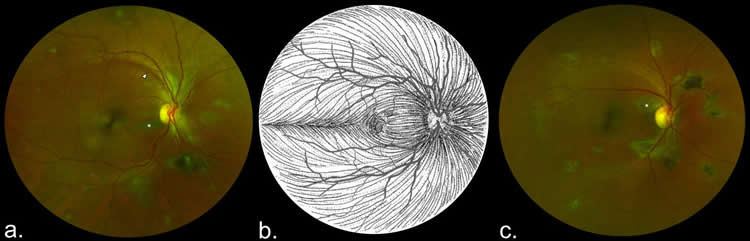
An eye team led by Dr Paul Steptoe, compared eye examinations of PES sufferers in Sierra Leone and the control population. A total of 82 Ebola survivors who had previously reported ocular symptoms and 105 unaffected controls from civilian and military personnel underwent ophthalmic examination, including widefield retinal imaging.
The results of the research, which has been published in the Emerging Infectious Diseases journal, shows that around 15% of Ebola survivors examined have a retinal scar that appears specific to the disease.
Reduced vision
Dr Steptoe, said: “The distribution of these retinal scars or lesions provides the first observational evidence that the virus enters the eye via the optic nerve to reach the retina in a similar way to West Nile Virus. Luckily, they appear to spare the central part of the eye so vision is preserved. Follow up studies are ongoing to assess for any potential recurrence of Ebola eye disease.
“Our study also provides preliminary evidence that in survivors with cataracts causing reduced vision but without evident active eye inflammation (uveitis), aqueous fluid analysis does not contain Ebola virus therefore enabling access to cataract surgery for survivors.”
Funding: This work was supported by The Dowager Countess Eleanor Peel Trust, Bayer Global Ophthalmology Awards Programme, Wellcome Trust ERAES Programme and the NIHR Health Protection Research Unit in Emerging and Zoonotic Infections at the University of Liverpool.
Source: Simon Wood – University of Liverpool
Image Source: NeuroscienceNews.com image is credited to University of Liverpool.
Original Research: Full open access research for “Novel Retinal Lesion in Ebola Survivors, Sierra Leone, 2016” by Paul J. Steptoe, Janet T. Scott, Julia M. Baxter, Craig K. Parkes, Rahul Dwivedi, Gabriela Czanner, Matthew J. Vandy, Fayiah Momorie, Alimamy D. Fornah, Patrick Komba, Jade Richards, Foday Sahr, Nicholas A.V. Beare, and Malcolm G. Semple in Emerging Infectious Diseases. Published online February 23 2017 doi:10.3201/eid2307.161608
[cbtabs][cbtab title=”MLA”]University of Liverpool “Ebola Survivors Have a ‘Unique’ Retinal Scar.” NeuroscienceNews. NeuroscienceNews, 16 May 2017.
<https://neurosciencenews.com/ebola-retina-scar-6696/>.[/cbtab][cbtab title=”APA”]University of Liverpool (2017, May 16). Ebola Survivors Have a ‘Unique’ Retinal Scar. NeuroscienceNew. Retrieved May 16, 2017 from https://neurosciencenews.com/ebola-retina-scar-6696/[/cbtab][cbtab title=”Chicago”]University of Liverpool “Ebola Survivors Have a ‘Unique’ Retinal Scar.” https://neurosciencenews.com/ebola-retina-scar-6696/ (accessed May 16, 2017).[/cbtab][/cbtabs]
Abstract
Novel Retinal Lesion in Ebola Survivors, Sierra Leone, 2016
We conducted a case–control study in Freetown, Sierra Leone, to investigate ocular signs in Ebola virus disease (EVD) survivors. A total of 82 EVD survivors with ocular symptoms and 105 controls from asymptomatic civilian and military personnel and symptomatic eye clinic attendees underwent ophthalmic examination, including widefield retinal imaging. Snellen visual acuity was <6/7.5 in 75.6% (97.5% CI 63%–85.7%) of EVD survivors and 75.5% (97.5% CI 59.1%–87.9%) of controls. Unilateral white cataracts were present in 7.4% (97.5% CI 2.4%–16.7%) of EVD survivors and no controls. Aqueous humor from 2 EVD survivors with cataract but no anterior chamber inflammation were PCR-negative for Zaire Ebola virus, permitting cataract surgery. A novel retinal lesion following the anatomic distribution of the optic nerve axons occurred in 14.6% (97.5% CI 7.1%–25.6%) of EVD survivors and no controls, suggesting neuronal transmission as a route of ocular entry. "Novel Retinal Lesion in Ebola Survivors, Sierra Leone, 2016” by Paul J. Steptoe, Janet T. Scott, Julia M. Baxter, Craig K. Parkes, Rahul Dwivedi, Gabriela Czanner, Matthew J. Vandy, Fayiah Momorie, Alimamy D. Fornah, Patrick Komba, Jade Richards, Foday Sahr, Nicholas A.V. Beare, and Malcolm G. Semple in Emerging Infectious Diseases. Published online February 23 2017 doi:10.3201/eid2307.161608


