


Using MRI, neuroscientists at Georgetown University Medical Center found significant differences in brain anatomy when comparing men and women with dyslexia to their non-dyslexic control groups, suggesting that the disorder may have a different brain-based manifestation based on sex.
Their study, investigating dyslexia in both males and females,is the first to directly compare brain anatomy of females with and without dyslexia (in children and adults). Their findings were published online in the journal Brain Structure and Function.
Because dyslexia is two to three times more prevalent in males compared with females, “females have been overlooked,” says senior author Guinevere Eden, PhD, director for the Center for the Study of Learning and past-president of the International Dyslexia Association.
“It has been assumed that results of studies conducted in men are generalizable to both sexes. But our research suggests that researchers need to tackle dyslexia in each sex separately to address questions about its origin and potentially, treatment,” Eden says.
Previous work outside of dyslexia demonstrates that male and female brains are different in general, adds the study’s lead author, Tanya Evans, PhD.
“There is sex-specific variance in brain anatomy and females tend to use both hemispheres for language tasks, while males just the left,” Evans says. “It is also known that sex hormones are related to brain anatomy and that female sex hormones such as estrogen can be protective after brain injury, suggesting another avenue that might lead to the sex-specific findings reported in this study.”
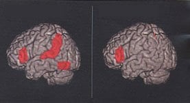
The study of 118 participants compared the brain structure of people with dyslexia to those without and was conducted separately in men, women, boys and girls. In the males, less gray matter volume is found in dyslexics in areas of the brain used to process language, consistent with previous work. In the females, less gray matter volume is found in dyslexics in areas involved in sensory and motor processing.
The results have important implications for understanding the origin of dyslexia and the relationship between language and sensory processing, says Evans.
Notes about this neuroanatomy and dyslexia research
The research funded by the Eunice Kennedy Shriver National Institute of Child Health and Human Development (P50HD40095 and R01HD05610701), by the National Center for Advancing Translational Sciences (UL1TR000101) and the National Science Foundation (SBE0541953 Science of Learning Center).
The authors report having no personal financial interests related to the study.
Contact: Karen Mallet – Georgetown University Medical Center
Source: Georgetown University Medical Center press release
Image Source: The dyslexia brain scan image is credited to NICHD/NIH and is available in the public domain.
Original Research: Abstract for “Sex-specific gray matter volume differences in females with developmental dyslexia” by Tanya M. Evans, D. Lynn Flowers, Eileen M. Napoliello and Guinevere F. Eden in Brain Structure and Function. Published online April 2013 doi: 10.1007/s00429-013-0552-4


