

NIH-funded study may guide deep brain stimulation therapies used for neurological disorders.
Scientists showed that they could alter brain activity of rats and either wake them up or put them in an unconscious state by changing the firing rates of neurons in the central thalamus, a region known to regulate arousal. The study, published in eLIFE, was partially funded by the National Institutes of Health.
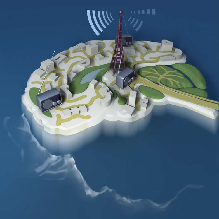
“Our results suggest the central thalamus works like a radio dial that tunes the brain to different states of activity and arousal,” said Jin Hyung Lee, Ph.D., assistant professor of neurology, neurosurgery and bioengineering at Stanford University, and a senior author of the study.
Located deep inside the brain the thalamus acts as a relay station sending neural signals from the body to the cortex. Damage to neurons in the central part of the thalamus may lead to problems with sleep, attention, and memory. Previous studies suggested that stimulation of thalamic neurons may awaken patients who have suffered a traumatic brain injury from minimally conscious states.
Dr. Lee’s team flashed laser pulses onto light sensitive central thalamic neurons of sleeping rats, which caused the cells to fire. High frequency stimulation of 40 or 100 pulses per second woke the rats. In contrast, low frequency stimulation of 10 pulses per second sent the rats into a state reminiscent of absence seizures that caused them to stiffen and stare before returning to sleep.
“This study takes a big step towards understanding the brain circuitry that controls sleep and arousal,” Yejun (Janet) He, Ph.D., program director at NIH’s National Institute of Neurological Disorders and Stroke (NINDS).
When the scientists used functional magnetic resonance imaging (fMRI) to scan brain activity, they saw that high and low frequency stimulation put the rats in completely different states of activity. Cortical brain areas where activity was elevated during high frequency stimulation became inhibited with low frequency stimulation. Electrical recordings confirmed the results. Neurons in the somatosensory cortex fired more during high frequency stimulation of the central thalamus and less during low frequency stimulation.
“Dr. Lee’s innovative work demonstrates the power of using imaging technologies to study the brain at work,” said Guoying Liu, Ph.D., a program director at the NIH’s National Institute of Biomedical Imaging and Bioengineering (NIBIB).
How can changing the firing rate of the same neurons in one region lead to different effects on the rest of the brain?
Further experiments suggested the different effects may be due to a unique firing pattern by inhibitory neurons in a neighboring brain region, the zona incerta, during low frequency stimulation. Cells in this brain region have been shown to send inhibitory signals to cells in the sensory cortex.
Electrical recordings showed that during low frequency stimulation of the central thalamus, zona incerta neurons fired in a spindle pattern that often occurs during sleep. In contrast, sleep spindles did not occur during high frequency stimulation. Moreover, when the scientists blocked the firing of the zona incerta neurons during low frequency stimulation of the central thalamus, the average activity of sensory cortex cells increased.
Although deep brain stimulation of the thalamus has shown promise as a treatment for traumatic brain injury, patients who have decreased levels of consciousness show slow progress through these treatments.
“We showed how the circuits of the brain can regulate arousal states,” said Dr. Lee. “We hope to use this knowledge to develop better treatments for brain injuries and other neurological disorders.”
Funding: This work was supported by grants from the NIH (NS087159, EB008738, MH087988); the National Science Foundation (CAREER Award, 1056008); the Okawa Foundation for Information and Telecommunications; the Alfred P. Sloan Foundation: the Mathers Charitable Foundation; Stanford Bio-X; James and Carrie Anderson Fund for Epilepsy Research; Littlefield Funds.
Source: Christopher G. Thomas – NIH/NINDS
Image Source: The image is credited to Lee lab, Stanford University, CA
Original Research: Full open access research for “Frequency-selective control of cortical and subcortical networks by central thalamus” by Jia Liu, Hyun Joo Lee, Andrew J Weitz, Zhongnan Fang, Peter Lin, ManKin Choy, Robert Fisher, Vadim Pinskiy, Alexander Tolpygo, Partha Mitra, Nicholas Schiff, and Jin Hyung Lee in eLife. Published online December 10 2015 doi:10.7554/eLife.09215
Abstract
Frequency-selective control of cortical and subcortical networks by central thalamus
Central thalamus plays a critical role in forebrain arousal and organized behavior. However, network-level mechanisms that link its activity to brain state remain enigmatic. Here, we combined optogenetics, fMRI, electrophysiology, and video-EEG monitoring to characterize the central thalamus-driven global brain networks responsible for switching brain state. 40 and 100 Hz stimulations of central thalamus caused widespread activation of forebrain, including frontal cortex, sensorimotor cortex, and striatum, and transitioned the brain to a state of arousal in asleep rats. In contrast, 10 Hz stimulation evoked significantly less activation of forebrain, inhibition of sensory cortex, and behavioral arrest. To investigate possible mechanisms underlying the frequency-dependent cortical inhibition, we performed recordings in zona incerta, where 10, but not 40, Hz stimulation evoked spindle-like oscillations. Importantly, suppressing incertal activity during 10 Hz central thalamus stimulation reduced the evoked cortical inhibition. These findings identify key brain-wide dynamics underlying central thalamus arousal regulation.
“Frequency-selective control of cortical and subcortical networks by central thalamus” by Jia Liu, Hyun Joo Lee, Andrew J Weitz, Zhongnan Fang, Peter Lin, ManKin Choy, Robert Fisher, Vadim Pinskiy, Alexander Tolpygo, Partha Mitra, Nicholas Schiff, and Jin Hyung Lee in eLife. Published online December 10 2015 doi:10.7554/eLife.09215


