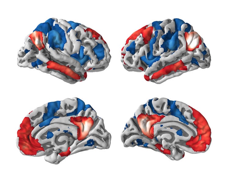
Over the past few years, a great amount of scientific research has shown that even when the brain is “at rest” it still works. The brains of healthy people are organized into regions displaying similar activity, called resting-state networks. There are two networks related to the perception of either the external world or internal thoughts. So far, much research on consciousness has focused on the activity within these networks, rather than how they communicate between each other.
An international research team has now investigated their interactions in different states of consciousness and has discovered that patients with severely impaired consciousness (disorders of consciousness, e.g. vegetative state/unresponsive wakefulness syndrome and minimally conscious state) show a pathological or uncontrolled communication between the two networks. Conversely, in patients who recovered from disorders of consciousness (patients known as emerging from minimally conscious state) the habitual interactions between networks are partially preserved, despite there being no difference in the connectivity within each network.
The study, published by the high-impact journal The Lancet Neurology, was led by Carol Di Perri, researcher at the Coma Science Group at Université de Liège (Belgium), and coordinated by Athena Demertzi, Steven Laureys (Coma Science Group) and Andrea Soddu from Western University (London, Ontario, Canada).
“Our findings cast light on the mechanisms underlying neural function necessary to emerge from impaired consciousness states and, more generally, on the importance of these network interactions in the emergence of higher cognitive functions,” says Laureys, Director, Coma Science Group and principal investigator at the GIGA (Interdisciplinary Cluster for Applied Geno-proteomics) of Liège. “Such results will have relevant impact in the clinical setting and may lead to possible new therapeutic options.”
According to Di Perri, previous research into disorders of consciousness predominantly focused on the connectivity breakdown within these networks, rather than their functional interactions.
“This study suggests that communication between networks is more important than connectivity within networks for cognitive functions necessary to emerge from disorders of consciousness,” says Di Perri.
Soddu, an assistant professor at Western’s Department of Physics and Astronomy and a member of the world-renowned Brain and Mind Institute, further explains, “This important discovery shows that the two neural networks interact in competing ways within the human brain, making patients emerging from minimally conscious state swing between the perception of the external world and their internal thoughts in an integrated manner.”
Source: Jeff Renaud – UWO
Image Source: The image is credited to the researchers.
Original Research: Abstract for “Neural correlates of consciousness in patients who have emerged from a minimally conscious state: a cross-sectional multimodal imaging study” by Carol Di Perri, Mohamed Ali Bahri, Enrico Amico, Aurore Thibaut, Lizette Heine, Georgios Antonopoulos, Vanessa Charland-Verville, Sarah Wannez, Francisco Gomez,, Roland Hustinx, Luaba Tshibanda, Athena Demertzi, Andrea Soddu, and Steven Laureys in Lancet Neurology. Published online April 27 2016 doi:10.1016/S1474-4422(16)00111-3
Abstract
Neural correlates of consciousness in patients who have emerged from a minimally conscious state: a cross-sectional multimodal imaging study
Background
Between pathologically impaired consciousness and normal consciousness exists a scarcely researched transition zone, referred to as emergence from minimally conscious state, in which patients regain the capacity for functional communication, object use, or both. We investigated neural correlates of consciousness in these patients compared with patients with disorders of consciousness and healthy controls, by multimodal imaging.
Methods
In this cross-sectional, multimodal imaging study, patients with unresponsive wakefulness syndrome, patients in a minimally conscious state, and patients who had emerged from a minimally conscious state, diagnosed with the Coma Recovery Scale–Revised, were recruited from the neurology department of the Centre Hospitalier Universitaire de Liège, Belgium. Key exclusion criteria were neuroimaging examination in an acute state, sedation or anaesthesia during scanning, large focal brain damage, motion parameters of more than 3 mm in translation and 3° in rotation, and suboptimal segmentation and normalisation. We acquired resting state functional and structural MRI data and 18F-fluorodeoxyglucose (FDG) PET data; we used seed-based functional MRI (fMRI) analysis to investigate positive default mode network connectivity (within-network correlations) and negative default mode network connectivity (between-network anticorrelations). We correlated FDG-PET brain metabolism with fMRI connectivity. We used voxel-based morphometry to test the effect of anatomical deformations on functional connectivity.
Findings
We recruited a convenience sample of 58 patients (21 [36%] with unresponsive wakefulness syndrome, 24 [41%] in a minimally conscious state, and 13 [22%] who had emerged from a minimally conscious state) and 35 healthy controls between Oct 1, 2009, and Oct 31, 2014. We detected consciousness-level-dependent increases (from unresponsive wakefulness syndrome, minimally conscious state, emergence from minimally conscious state, to healthy controls) for positive and negative default mode network connectivity, brain metabolism, and grey matter volume (p<0·05 false discovery rate corrected for multiple comparisons). Positive default mode network connectivity differed between patients and controls but not among patient groups (F test p<0·0001). Negative default mode network connectivity was only detected in healthy controls and in those who had emerged from a minimally conscious state; patients with unresponsive wakefulness syndrome or in a minimally conscious state showed pathological between-network positive connectivity (hyperconnectivity; F test p<0·0001). Brain metabolism correlated with positive default mode network connectivity (Spearman's r=0·50 [95% CI 0·26 to 0·61]; p<0·0001) and negative default mode network connectivity (Spearman's r=–0·52 [–0·35 to −0·67); p<0·0001). Grey matter volume did not differ between the studied groups (F test p=0·06).
Interpretation
Partial preservation of between-network anticorrelations, which are seemingly of neuronal origin and cannot be solely explained by morphological deformations, characterise patients who have emerged from a minimally conscious state. Conversely, patients with disorders of consciousness show pathological between-network correlations. Apart from a deeper understanding of the neural correlates of consciousness, these findings have clinical implications and might be particularly relevant for outcome prediction and could inspire new therapeutic options.
Funding
Belgian National Funds for Scientific Research (FNRS), European Commission, Natural Sciences and Engineering Research Council of Canada, James McDonnell Foundation, European Space Agency, Mind Science Foundation, French Speaking Community Concerted Research Action, Fondazione Europea di Ricerca Biomedica, University and University Hospital of Liège (Liège, Belgium), and University of Western Ontario (London, ON, Canada).
“Neural correlates of consciousness in patients who have emerged from a minimally conscious state: a cross-sectional multimodal imaging study” by Carol Di Perri, Mohamed Ali Bahri, Enrico Amico, Aurore Thibaut, Lizette Heine, Georgios Antonopoulos, Vanessa Charland-Verville, Sarah Wannez, Francisco Gomez,, Roland Hustinx, Luaba Tshibanda, Athena Demertzi, Andrea Soddu, and Steven Laureys in Lancet Neurology. Published online April 27 2016 doi:10.1016/S1474-4422(16)00111-3