

McLean scientists link activity in two brain regions to fear responses.
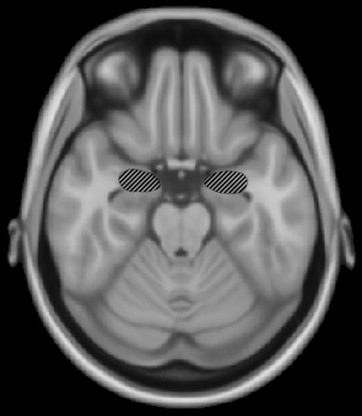
In a new paper published in the current issue of Neuron, Harvard Medical School researchers at McLean Hospital report that increased activity in the medial prefrontal cortex of the brain is linked to decreased activity in the amygdala, the portion of the brain where memories of frightening events are created.
According to study author Vadim Bolshakov, HMS professor of psychiatry at McLean, this finding could lead to better methods to prevent post-traumatic stress disorder (PTSD).
“A single exposure to something traumatic or scary can be enough to create a fear memory, causing someone to expect and be afraid in similar situations in the future,” said Bolshakov. “What we’re seeing is that we may one day be able to prevent those fear memories.”
Bolshakov and his colleagues tested their theory in mice. Divided into two groups, some were taught to fear an auditory stimulus while others were taught fear but had that memory extinguished. In the animals whose fear was extinguished, increased activation of the medial prefrontal cortex led to inhibition of the amygdala and significant decreases in fear responses.
“For example, if a sound ended with an extremely loud shriek, a subject would come to expect that scary noise at the end of the sound,” explained Bolshakov. “What we found was when we suppressed the fear memory by decreasing activity in the amygdala, the subjects were not afraid of the end of the auditory stimulus any longer.”
Bolshakov noted that this work could have important implications for the treatment of a number of conditions, including PTSD.
“While there is still a great deal of research that needs to be done before our work can be translated to clinical trials, what we are showing has the potential to ensure that individuals exposed to trauma were not haunted by the conditions surrounding their initial stressor.”
Notes about this neuroscience and fear research
The study was supported by Whitehall Foundation and NIH grants MH083011 and MH090464.
Contact: Scott O’Brien – McLean Hospital
Source: McLean Hospital press release
Image Source: The image is credited to Woutergroen and is in the public domain.
Original Research: Abstract for “Synaptic Encoding of Fear Extinction in mPFC-amygdala Circuits” by Jun-Hyeong Cho, Karl Deisseroth, and Vadim Y. Bolshakov in Neuron. Published online November 27 2013 doi:10.1016/j.neuron.2013.09.025


