Summary: A new study published in Science reveals glia are vital for nerve cell development in the brain.
Source: NYU.
A team of biologists has found an unexpected source for the brain’s development, a finding that offers new insights into the building of the nervous system.
The research, which appears in the journal Science, discovered that glia, a collection of non-neuronal cells that had long been regarded as passive support cells, in fact are vital to nerve-cell development in the brain.
“The results lead us to revise the often neuro-centric view of brain development to now appreciate the contributions for non-neuronal cells such as glia,” explains Vilaiwan Fernandes, a postdoctoral fellow in New York University’s Department of Biology and the study’s lead author. “Indeed, our study found that fundamental questions in brain development with regard to the timing, identity, and coordination of nerve cell birth can only be understood when the glial contribution is accounted for.”
The brain is made up of two broad cell types, nerve cells or neurons and glia, which are non-nerve cells that make up more than half the volume of the brain. Neurobiologists have tended to focus on the former because these are the cells that form networks that process information.
However, given the preponderance of glia in the brain’s cellular make-up, the NYU researchers hypothesized that they could play a fundamental part in brain development.
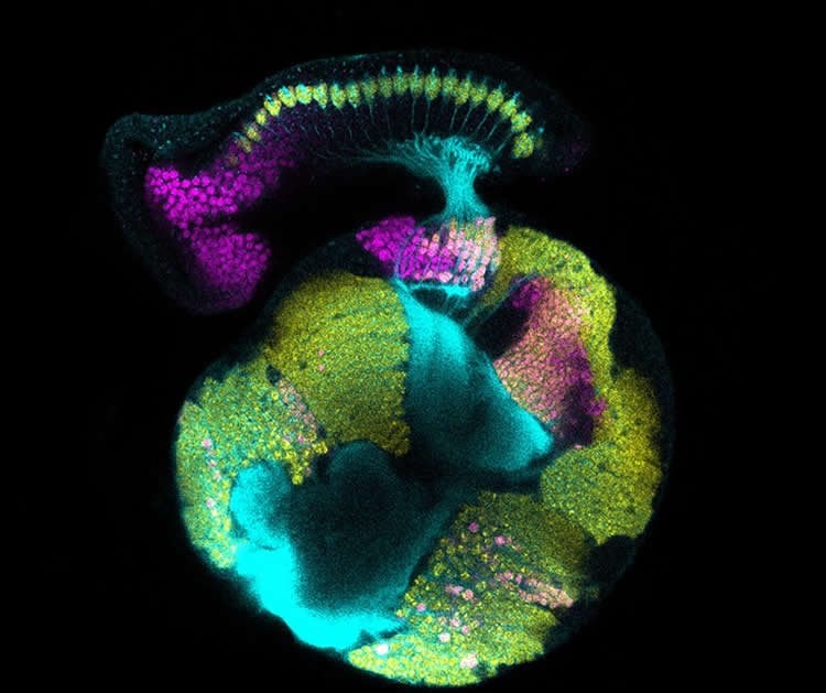
To explore this, they examined the visual system of the fruit fly. The species serves as a powerful model organism for this line of study because its visual system, like the one in humans, holds repeated mini-circuits that detect and process light over the entire visual field.
This dynamic is of particular interest to scientists because, as the brain develops, it must coordinate the increase of neurons in the retina with other neurons in distant regions of the brain.
In their study, the NYU researchers found that the coordination of nerve-cell development is achieved through a population of glia, which relay cues from the retina to the brain to make cells in the brain become nerve cells.
“By acting as a signaling intermediary, glia exert precise control over not only when and where a neuron is born, but also the type of neuron it will develop into,” notes NYU Biology Professor Claude Desplan, the paper’s senior author.
Funding: The research was supported, in part, by a grant from the National Institutes of Health (EY13012).
Source: James Devitt – NYU
Image Source: NeuroscienceNews.com image is credited to Vilaiwan M Fernandes, Desplan Lab, NYU’s Department of Biology.
Original Research: Abstract for “Glia relay differentiation cues to coordinate neuronal development in Drosophila” by Vilaiwan M. Fernandes, Zhenqing Chen, Anthony M. Rossi, Jaqueline Zipfel, and Claude Desplan in Science. Published online September 1 2017 doi:10.1126/science.aan3174
[cbtabs][cbtab title=”MLA”]NYU “New Source For Brain’s Development Discovered.” NeuroscienceNews. NeuroscienceNews, 3 September 2017.
<https://neurosciencenews.com/brain-development-neurobiology-7401/>.[/cbtab][cbtab title=”APA”]NYU (2017, September 3). New Source For Brain’s Development Discovered. NeuroscienceNew. Retrieved September 3, 2017 from https://neurosciencenews.com/brain-development-neurobiology-7401/[/cbtab][cbtab title=”Chicago”]NYU “New Source For Brain’s Development Discovered.” https://neurosciencenews.com/brain-development-neurobiology-7401/ (accessed September 3, 2017).[/cbtab][/cbtabs]
Abstract
Glia relay differentiation cues to coordinate neuronal development in Drosophila
Neuronal birth and specification must be coordinated across the developing brain to generate the neurons that constitute neural circuits. We used the Drosophila visual system to investigate how development is coordinated to establish retinotopy, a feature of all visual systems. Photoreceptors achieve retinotopy by inducing their target field in the optic lobe, the lamina neurons, with a secreted differentiation cue, epidermal growth factor (EGF). We find that communication between photoreceptors and lamina cells requires a signaling relay through glia. In response to photoreceptor-EGF, glia produce insulin-like peptides, which induce lamina neuronal differentiation. Our study identifies a role for glia in coordinating neuronal development across distinct brain regions, thus reconciling the timing of column assembly with that of delayed differentiation, as well as the spatiotemporal pattern of lamina neuron differentiation.
“Glia relay differentiation cues to coordinate neuronal development in Drosophila” by Vilaiwan M. Fernandes, Zhenqing Chen, Anthony M. Rossi, Jaqueline Zipfel, and Claude Desplan in Science. Published online September 1 2017 doi:10.1126/science.aan3174