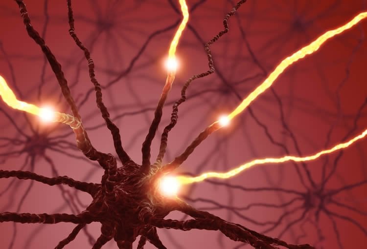
Specific regions of the brain are specialized in recognizing bodies of animals and human beings. By measuring the electrical activity per cell, scientists from KU Leuven, Belgium, and the University of Glasgow have shown that the individual brain cells in these areas do different things. Their response to specific contours or body shapes is very selective.
Facial recognition has already been the subject of much research. But what happens when we cannot recognize an animal or a human being on the basis of a face, but only have other body parts to go on? The mechanism behind this recognition process is uncharted territory for neuroscientists, says Professor Rufin Vogels of the KU Leuven Laboratory for Neuro- and Psychophysiology.
“Previous research in monkeys has shown that small areas in the temporal lobes – the parts of the brain near the temples – are activated when the monkeys look at bodies instead of objects or faces. Brain scans tell us that these regions of the brain correspond to the ones activated in human beings. But that only tells us which regions are active, not which information about bodies is passed on by their cells.”
The KU Leuven scientists measured the electrical activity of individual cells in one of the brain areas responsible for body recognition in rhesus monkeys. Vogels explains: “We showed the monkeys images of bodies of both animals and human beings, while covering up specific body parts. We usually left out the faces, because other brain cells are responsible for recognizing those. With a method developed at the University of Glasgow, we then examined each cell to see which body parts activated it.”

“We found that these individual cells in themselves are not body detectors: they respond to specific, frequently found characteristics of bodies, such as the curve of an elbow or a part of a leg. And they divide the work. Each cell has its own speciality and screens the incoming information for certain characteristics. The brain recognizes a body by combining all the pieces of the puzzle: all the bits of information registered by the different cells in a particular region of the brain. The brain cells, in other words, collaborate to solve the puzzle.”
In their next project, the scientists want to expand their study to include a number of other brain areas that are also active for body recognition. “If we improve our understanding of how our brains recognize bodies, we can use that knowledge for disorders whereby that particular mechanism is disrupted, such as autism, or for the development of facial-recognition software.”
Source: Adam Phillips – KU Leuven
Image Source: The image is adapted from the KU Leuven press release.
Original Research: Abstract for “Stimulus features coded by single neurons of a macaque body category selective patch” by Ivo D. Popivanov, Philippe G. Schyns, and Rufin Vogels in PNAS. Published online March 18 2016 doi:10.1073/pnas.1520371113
Abstract
Stimulus features coded by single neurons of a macaque body category selective patch
Body category-selective regions of the primate temporal cortex respond to images of bodies, but it is unclear which fragments of such images drive single neurons’ responses in these regions. Here we applied the Bubbles technique to the responses of single macaque middle superior temporal sulcus (midSTS) body patch neurons to reveal the image fragments the neurons respond to. We found that local image fragments such as extremities (limbs), curved boundaries, and parts of the torso drove the large majority of neurons. Bubbles revealed the whole body in only a few neurons. Neurons coded the features in a manner that was tolerant to translation and scale changes. Most image fragments were excitatory but for a few neurons both inhibitory and excitatory fragments (opponent coding) were present in the same image. The fragments we reveal here in the body patch with Bubbles differ from those suggested in previous studies of face-selective neurons in face patches. Together, our data indicate that the majority of body patch neurons respond to local image fragments that occur frequently, but not exclusively, in bodies, with a coding that is tolerant to translation and scale. Overall, the data suggest that the body category selectivity of the midSTS body patch depends more on the feature statistics of bodies (e.g., extensions occur more frequently in bodies) than on semantics (bodies as an abstract category).
“Stimulus features coded by single neurons of a macaque body category selective patch” by Ivo D. Popivanov, Philippe G. Schyns, and Rufin Vogels in PNAS. Published online March 18 2016 doi:10.1073/pnas.1520371113


