Summary: A new 3D imaging method allows researchers to measure brain growth and folding patterns in a baby’s brain during the third trimester of pregnancy.
Source: WUSTL.
During the third trimester, a baby’s brain undergoes rapid development in utero. The cerebral cortex dramatically expands its surface area and begins to fold. Previous work suggests that this quick and very vital growth is an individualized process, with details varying infant to infant.
Research from a collaborative team at Washington University in St. Louis tested a new, 3-D method that could lead to new diagnostic tools that will precisely measure the third-trimester growth and folding patterns of a baby’s brain.
The findings, published online March 5 in PNAS, could help to sound an early alarm on developmental disorders in premature infants that could affect them later in life.
“One of the things that’s really interesting about people’s brains is that they are so different, yet so similar,” said Philip Bayly, the Lilyan & E. Lisle Hughes Professor of Mechanical Engineering at the School of Engineering & Applied Science. “We all have the same components, but our brain folds are like fingerprints: Everyone has a different pattern. Understanding the mechanical process of folding — when it occurs — might be a way to detect problems for brain development down the road.”
Working with collaborators at Washington University School of Medicine in St. Louis, engineering doctoral student Kara Garcia accessed magnetic resonance 3-D brain images from 30 pre-term infants scanned by Christopher Smyser, MD, associate professor of neurology, and his pediatric neuroimaging team. The babies were scanned two to four times each during the period of rapid brain expansion, which typically happens at 28 to 30 weeks.
Using a new computer algorithm, Bayly, Garcia and their colleagues — including faculty at Imperial College and King’s College in London — obtained accurate point-to-point correspondence between younger and older cortical reconstructions of the same infant. From each pair of surfaces, the team calculated precise maps of cortical expansion. Then, using a minimum energy approach to compare brain surfaces at different times, researchers picked up on subtle differences in the babies’ brain folding patterns.
“The minimum energy approach is the one that’s most likely from a physical standpoint,” Bayly said. “When we obtain surfaces from MR images, we don’t know which points on the older surface correspond with which points on the younger surface. We reasoned, that since nature is efficient, the most likely correspondence is the one that produces the best match between surface landmarks, while at the same time, minimizing how much the brain would have to distort while it is growing.
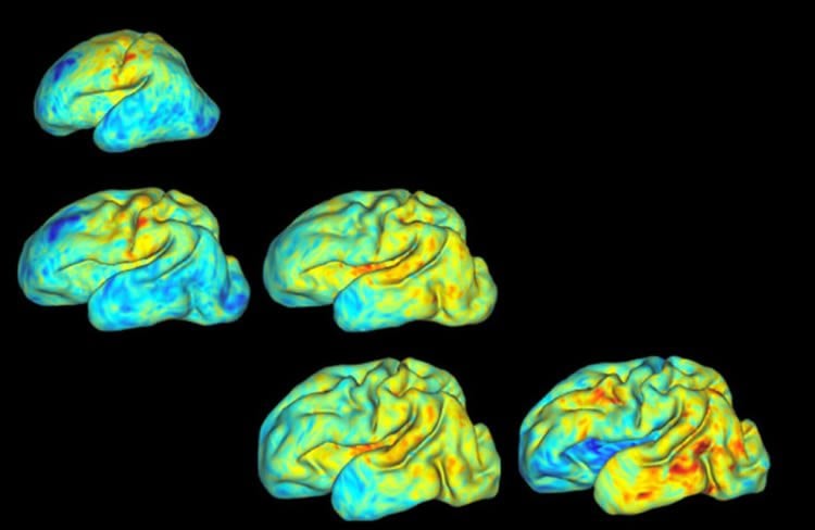
“When you use this minimum energy approach, you get rid of a lot of noise in the analysis, and what emerged were these subtle patterns of growth that were previously hidden in the data. Not only do we have a better picture of these developmental processes in general, but doctors should hopefully be able to assess individual patients, take a look at their pattern of brain development, and figure out how it’s tracking.”
It’s a measurement tool that could prove invaluable in places such as neonatal intensive care units, where preemies face a variety of challenges. Understanding an individual’s precise pattern of brain development also could assist physicians trying to make a diagnosis later in a patient’s life.
“You do also find folding abnormalities in populations that have cognitive issues later in life, including autism and schizophrenia,” Bayly said. “It’s possible, if medical researchers understand better the folding process and what goes on wrong or differently, then they can understand a little bit more about what causes these problems.”
Funding: This work was supported by National Institute of Health grants R01 NS055951, R01 NS070918, T32 EB018266, K02 NS089852, K23 MH105179, UL1 TR000448
Source: Erika Ebsworth-Goold – WUSTL
Publisher: Organized by NeuroscienceNews.com.
Image Source: NeuroscienceNews.com image is credited to Bayly Lab.
Original Research: Abstract in PNAS.
doi:10.1073/pnas.1715451115
[cbtabs][cbtab title=”MLA”]WUSTL “3D Mapping Babies’ Brains.” NeuroscienceNews. NeuroscienceNews, 8 March 2018.
<https://neurosciencenews.com/baby-brain-mapping-8610/>.[/cbtab][cbtab title=”APA”]WUSTL (2018, March 8). 3D Mapping Babies’ Brains. NeuroscienceNews. Retrieved March 8, 2018 from https://neurosciencenews.com/baby-brain-mapping-8610/[/cbtab][cbtab title=”Chicago”]WUSTL “3D Mapping Babies’ Brains.” https://neurosciencenews.com/baby-brain-mapping-8610/ (accessed March 8, 2018).[/cbtab][/cbtabs]
Abstract
Dynamic patterns of cortical expansion during folding of the preterm human brain
During the third trimester of human brain development, the cerebral cortex undergoes dramatic surface expansion and folding. Physical models suggest that relatively rapid growth of the cortical gray matter helps drive this folding, and structural data suggest that growth may vary in both space (by region on the cortical surface) and time. In this study, we propose a unique method to estimate local growth from sequential cortical reconstructions. Using anatomically constrained multimodal surface matching (aMSM), we obtain accurate, physically guided point correspondence between younger and older cortical reconstructions of the same individual. From each pair of surfaces, we calculate continuous, smooth maps of cortical expansion with unprecedented precision. By considering 30 preterm infants scanned two to four times during the period of rapid cortical expansion (28–38 wk postmenstrual age), we observe significant regional differences in growth across the cortical surface that are consistent with the emergence of new folds. Furthermore, these growth patterns shift over the course of development, with noninjured subjects following a highly consistent trajectory. This information provides a detailed picture of dynamic changes in cortical growth, connecting what is known about patterns of development at the microscopic (cellular) and macroscopic (folding) scales. Since our method provides specific growth maps for individual brains, we are also able to detect alterations due to injury. This fully automated surface analysis, based on tools freely available to the brain-mapping community, may also serve as a useful approach for future studies of abnormal growth due to genetic disorders, injury, or other environmental variables.