Researchers from the University of Houston have analyzed brain activity data collected from more than 400 people who viewed an exhibit at the Menil Collection, offering evidence that useable brain data can be collected outside of a controlled laboratory setting. They also reported the first real-world demonstration of what happens in the brain as people observe artwork.
“You can do testing in the lab, but it’s very artificial,” said Jose Luis Contreras-Vidal, Hugh Roy and Lillie Cranz Cullen Distinguished Professor of electrical and computer engineering at UH. “We were looking at how to measure brain activity in action and in context.”
The researchers reported their findings in the journal Frontiers in Human Neuroscience. In addition to Contreras-Vidal, the research team included Kimberly Kontson and Eugene Civillico, scientists with the U.S. Food and Drug Administration; artist Dario Robleto; Menil curator Michelle White, and Murad Megjhani, Justin Brantley, Jesus Cruz-Garza and Sho Nakagome, all of whom work in the UH Laboratory for Non-Invasive Brain Machine Interfaces.
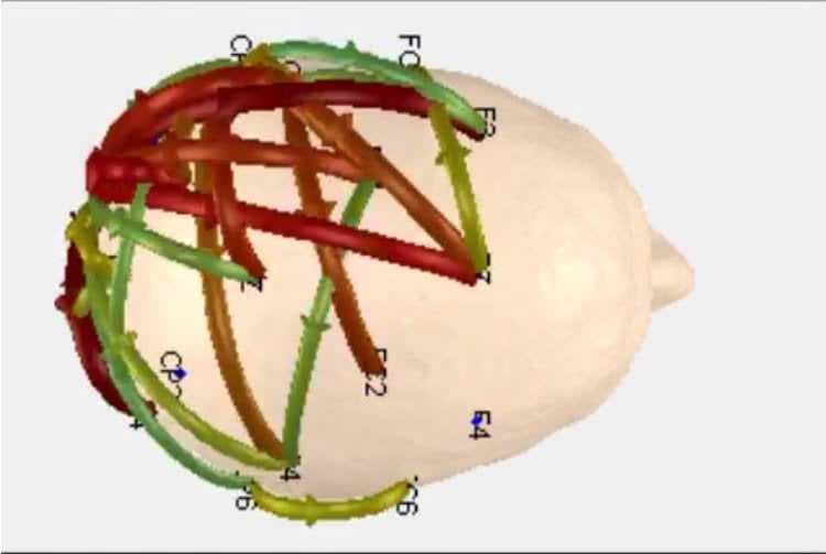
The research found significant increases in functional, or task-related, connectivity in localized brain networks when the subjects viewed art they considered aesthetically pleasing, compared with baseline readings. They found differences both between men and women and between the youngest and oldest subjects.
“The direction of signal flow showed early recruitment of broad posterior [visual] areas followed by focal anterior activation,” they wrote. “Significant differences in the strength of connections were also observed across age and gender. This work provides evidence that EEG [electroencephalogram], deployed on freely behaving subjects, can detect selective signal flow in neural networks, identify significant differences between subject groups, and report with greater-than-chance accuracy the complexity of a subject’s visual percept of aesthetically pleasing art.”
Kontson, a biomedical research fellow at the FDA who led the research during a post-doctoral fellowship, said researchers started with three questions: Can useable brain data be collected in an uncontrolled setting? How well do different models of EEG headsets perform? Is it possible to collect substantial amounts of data relatively quickly?
EEG headsets are considered medical devices if intended for use in the diagnosis of disease or other conditions. Kontson said the FDA is interested in the potential use of large and complex data sets – “big data” – for regulatory decision-making.

Data was collected from 431 people as they viewed Robleto’s solo show at the Menil Collection in Houston, “The Boundary of Life Is Quietly Crossed,” a sculptural installation that included both visual and aural representations of the heart. Researchers categorized each piece as either complex or moderate; they also asked each participant to face a blank wall for one minute before entering the exhibit in order to obtain baseline data.
The Frontiers in Human Neuroscience paper is based on data from 20 people who wore a reference gel-based EEG headset; Contreras-Vidal said findings from those who wore one of four models of dry-electrode headsets – easier to use in public, as they require little preparation or instruction – will be reported later, as will an analysis on data collected with the dry headsets.
The initial results allowed researchers to predict from the brain activity with 55 percent accuracy whether the participant was looking at a complex piece of art, one categorized as moderately complex or a blank wall. That compares to 33 percent accuracy for random prediction.
The knowledge could have varying applications. Much of Contreras-Vidal’s recent work centers on using brain activity to help people with disabilities use bionic hands or to regain movement by “walking” in exoskeletons powered by their own thoughts. He sees this research with artists and museum-goers – a related project collects brain activity from dancers, visual artists, musicians and writers – as potentially leading to technologies that can restore sensory processing in people with neurological impairments.
Artists and museum curators could use the findings to learn more about how museum displays affect the way people move through and react to an exhibit, which works are preferred by museum-goers and other information, Contreras-Vidal.
But he doesn’t expect the research to produce a how-to on creating art.
“I don’t think we will understand the mystery (of how art is created),” he said. “The conception of art is a very individual process, built on the artist’s experiences, skills, memories, values and drives. But we will know what happens in the brain. We might find that there are people who are very attuned to visual art, or to music, or poetry, and there might be an underlying common neural network. If we know that, we could optimize the delivery of art for therapy, for teaching.”
Source: Jeannie Kever – University of Houston
Image Credit: The image is adapted from the UHmultimedia video
Video Source: The video is available at the UHmultimedia YouTube page
Original Research: Abstract for “Your Brain on Art: Emergent cortical dynamics during aesthetic experiences” by Kimberly Kontson, Murad Megjhani, Justin A. Brantley, Jesus G. Cruz-Garza, Sho Nakagome, Dario Robleto, Michelle White, Eugene Civillico and Jose L. Contreras-Vidal in Frontiers in Human Neuroscience. Published online Novemeber 2 2015 doi:10.3389/fnhum.2015.00626
Abstract
‘Your Brain on Art’: Emergent cortical dynamics during aesthetic experiences
The brain response to conceptual art was studied with mobile electroencephalography (EEG) to examine the neural basis of aesthetic experiences. In contrast to most studies of perceptual phenomena, participants were moving and thinking freely as they viewed the exhibit The Boundary of Life is Quietly Crossed by Dario Robleto at the Menil Collection-Houston. The brain activity of over 400 subjects was recorded using dry-electrode and one reference gel-based EEG systems over a period of 3 months. Here, we report initial findings based on the reference system. EEG segments corresponding to each art piece were grouped into one of three classes (complex, moderate, and baseline) based on analysis of a digital image of each piece. Time, frequency, and wavelet features extracted from EEG were used to classify patterns associated with viewing art, and ranked based on their relevance for classification. The maximum classification accuracy was 55% (chance = 33%) with delta and gamma features the most relevant for classification. Functional analysis revealed a significant increase in connection strength in localized brain networks while subjects viewed the most aesthetically pleasing art compared to viewing a blank wall. The direction of signal flow showed early recruitment of broad posterior areas followed by focal anterior activation. Significant differences in the strength of connections were also observed across age and gender. This work provides evidence that EEG, deployed on freely behaving subjects, can detect selective signal flow in neural networks, identify significant differences between subject groups, and report with greater-than-chance accuracy the complexity of a subject’s visual percept of aesthetically pleasing art. Our approach, which allows acquisition of neural activity ‘in action and context’, could lead to understanding of how the brain integrates sensory input and its ongoing internal state to produce the phenomenon which we term aesthetic experience.
“Your Brain on Art: Emergent cortical dynamics during aesthetic experiences” by Kimberly Kontson, Murad Megjhani, Justin A. Brantley, Jesus G. Cruz-Garza, Sho Nakagome, Dario Robleto, Michelle White, Eugene Civillico and Jose L. Contreras-Vidal in Frontiers in Human Neuroscience. Published online Novemeber 2 2015 doi:10.3389/fnhum.2015.00626


