

Summary: Two new studies strengthen the link between gum disease and the development of Alzheimer’s disease. The studies focus on the interactions between the gingipains enzyme interacts with the Tau protein, and how the gingipains enzyme contributes to the development of amyloid-beta.
Source: UCLan
Researchers at the School of Dentistry, University of Central Lancashire (UCLan) were the first to report the link between gum disease and Alzheimer’s disease.
Now two new studies from the same research group at the School of Dentistry demonstrate that progress is being made in making much stronger connections between gum disease in the mouth and deteriorating brain function.
The studies are incredibly timely, as September is the World Alzheimer’s month and September 21 is World Alzheimer’s day. Marking this important awareness day, the two new studies—published in the Journal of Alzheimer’s Disease and the Journal of Alzheimer’s Disease Reports, respectively—give a better understanding of the defining Alzheimer’s disease lesions on the brain: technically known as amyloid-beta plaques and neurofibrillary tangles.
Alzheimer’s disease is the most common type of dementia, progressively causing deterioration of memory, thinking skills and the ability to communicate. The exact cause of this disease is not yet fully understood, which means it’s a difficult disease to prevent and treat.
It has been previously shown that the Porphyromonas gingivalis bacterium which destroys gum tissue—and the enzyme which it produces, known as gingipains—are specifically linked to Alzheimer’s disease, after both were discovered in the brain tissue of those suffering from the disease. These new studies go a step further in exploring how gum disease and its bacterial proteins can potentially contribute to the formation of lesions on the brain.
The first study, to be published in the Journal of Alzheimer’s Disease, shows that nerve cells in the brain contain a type of protein called tau. When tau meets the gingipains enzyme, the tau released from the nerve cell. Once freed, tau physically changes, in the form of coils and non-coiling filaments. These filaments of tau then re-attach to the nerve cell and become incorporated into the lesion known as neurofibrillary tangles. These ultimately kill the nerve cells.
What this means is that once a nerve cell dies and the free tau protein leaks into the brain, the tau may attach itself to healthy neighboring nerve cells, repeating the process and leading to further damage to the brain as the disease spreads.
The second study, published in the Journal of Alzheimer’s Disease Reports, looks at the way the gingipains enzyme, released by the bacterium, can contribute to the formation of amyloid-beta plaques—another of the lesions, alongside the tangles, which form on the brains of those suffering from Alzheimer’s.
These studies are small steps in the fight against Alzheimer’s, but the results are significant in understanding the role of gingipains and how fundamental they are to lesion formation. It is hoped that studies such as these will help with developing future treatments.
Shalini Kanagasingam, Specialist Endodontist and Senior Clinical Lecturer at UCLan, who led the study, (supervised by Dr. Sim K. Singhrao) said, “What this kind of research proves is the importance of our oral health. Look out for early signs of gum disease such as bleeding gums when brushing or more advanced signs like movement or drifting of the teeth.
“Don’t delay or skip your dental check-ups. Your dentist will be able to help advise you on how to effectively remove plaque and tartar from your teeth, which harbor the bacterium that we have identified as a risk factor for Alzheimer’s.
“These studies highlight the key message that a healthy mouth is important for maintaining a healthy body and mind.”
About this gum disease and Alzheimer’s disease research news
Author: Press Office
Contact: UCLan
Source: Press Office – UCLan
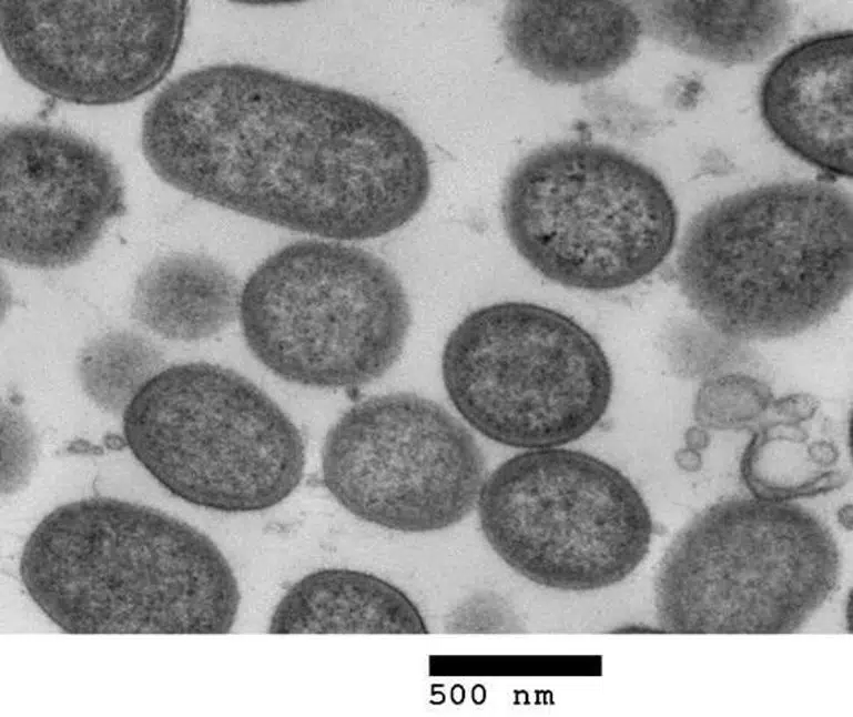
Image: The image is credited to UCLan
Original Research: Closed access.
“Antimicrobial, Polarizing Light, and Paired Helical Filament Properties of Fragmented Tau Peptides of Selected Putative Gingipains” by Shalini Kanagasingam et al. Journal of Alzheimer’s Disease
Open access.
“Porphyromonas gingivalis Conditioned Medium Induces Amyloidogenic Processing of the Amyloid-β Protein Precursor upon in vitro Infection of SH-SY5Y Cells” by Shalini Kanagasingam et al. Journal of Alzheimer’s Disease Reports
Abstract
Antimicrobial, Polarizing Light, and Paired Helical Filament Properties of Fragmented Tau Peptides of Selected Putative Gingipains
Background: Tau is an established substrate for gingipains secreted by Porphyromonas gingivalis. Hyperphosphorylation of tau and neurofibrillary tangle (NFT) formation is a defining lesion of Alzheimer’s disease (AD) where NFT distribution is related to Braak stage and disease severity.
Objective: To assess gingipains’-fragmented tau peptides for their antimicrobial properties and for the likelihood of paired helical/straight filament (PHF/SF) formation with implications for the NFT lesion.
Methods: Seven non-phosphorylated (A-G) and three phosphorylated (A-C) tau peptides, were tested for antimicrobial properties against P. gingivalis. Polarizing light properties were determined using Congo Red staining. Secondary and tertiary structures of peptides B-F were determined using transmission electron microscopy (TEM) and circular dichroism (CD) was undertaken for the soluble peptides A in phosphorylated and non-phosphorylated states.
Results: Phosphorylated tau peptide A displayed a significant effect against planktonic P. gingivalis. The CD results demonstrated that both peptides A, in phosphorylated and non-phosphorylated states, in aqueous solution, adopted mainly β-type structures. Non-phosphorylated peptides B-F and phosphorylated peptides B-C were insoluble and fibrillar under the TEM. The secondary and tertiary structures of the non-phosphorylated peptide B demonstrated fewer helical twists, whereas peptide C displayed significantly more helical twists along the whole fiber(s) length following its phosphorylation.
Conclusion: Phosphorylated peptide A reduced P. gingivalis viability. CD spectroscopy demonstrated the phosphorylated and the non-phosphorylated peptide A predominantly formed from β-sheet structures in aqueous solution with potential antimicrobial activity. Phosphorylation of tau peptides physically changed their tertiary structure into PHFs with potential for self-aggregation and binding to the NFT lesion.
Abstract
Porphyromonas gingivalis Conditioned Medium Induces Amyloidogenic Processing of the Amyloid-β Protein Precursor upon in vitro Infection of SH-SY5Y Cells
Background:
Cleavage of the amyloid-β protein precursor (AβPP) mediated by host secretase enzymes, releases several fragments including amyloid-β (Aβ40 and Aβ42).
Objective:
To determine if Porphyromonas gingivalis conditioned medium cleaved AβPP to release Aβ40 and Aβ42.
Methods:
The SH-SY5Y cell line was challenged, in vitro, with P. gingivalis (Pg381) conditioned medium in the presence/absence of cytokines. The cells and their supernatants were assessed for AβPP cleavage fragments by immunoblotting and transmission electron microscopy.
Results:
Western blotting of the cell lysates with the anti-AβPP C-terminal antibody demonstrated variable molecular weight bands corresponding to full length and fragmented AβPP in lanes treated with the following factors: Tryptic soy broth (TSB), Pg381, IL-6, Pg381 + IL-1β, and Pg381 + TNF-α. The low molecular weight bands corresponding to the C99 dimerized fragment were observed in the Pg381 and interlukin-6 (IL-6) treated groups and were significantly more intense in the presence of Pg381 with either IL-6 or TNF-α. Bands corresponding to the dimerized C83 fragment were observed with cells treated with TNF-α alone and with Pg381 combined with IL-1β or IL-6 or TNF-α. The anti-Aβ antibody detected statistically significant Aβ40 and Aβ42, levels when these two Aβ species were pooled across test samples and compared to the untreated group. Electron microscopic examination of the supernatants demonstrated insoluble Aβ40 and Aβ42.
Conclusion:
These observations strongly imply that AβPP is an infection responsive protein cleaved via the amyloidogenic pathway on exposure to conditioned medium and in the presence of pro-inflammatory mediators.


