Summary: Disruptions in brain activity during a common form of childhood seizures are more global than researchers previously believed, a new study reports.
Source: Yale.
Scientists believed that absence seizures — the brief loss of consciousness often mistaken for day-dreaming — was caused by a localized disruption of brain activity. A new Yale study finds the entire brain is involved in this common form of childhood epilepsy that causes kids to “blank out” for 10 seconds or more at a time.
“These seizures significantly affect school performance, social interactions and can also pose safety risks,” said Dr. Hal Blumenfeld, the Mark Loughridge and Michele Williams Professor of Neurology and senior author of the study appearing Nov. 7 in the journal Lancet Neurology.
Seizures typically last less than 10 seconds but can also last longer and be accompanied by movements such as blinking, chewing or hand gestures. In severe cases, these seizures can happen hundreds of times a day.
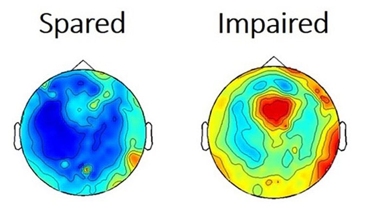
The Yale team collected brain electrical signaling and fMRI data from 39 children undergoing absence seizures and found disruptions were global, not localized. Also, they found that in more severe seizures, disruptions began even before symptoms of seizures began.
“Understanding impaired consciousness in childhood absence epilepsy can also improve understanding of other disorders of consciousness including head trauma, coma and stroke,” Blumenfeld said.
Funding: National Institutes of Health, National Institute of Neurological Disorders and Stroke, National Center for Advancing Translational Science, the Loughridge Williams Foundation, and the Betsy and Jonathan Blattmachr Family.
Source: Bill Hathaway – Yale
Image Source: This NeuroscienceNews.com image is credited to Blumenfeld lab.
Original Research: Abstract for “Impaired consciousness in patients with absence seizures investigated by functional MRI, EEG, and behavioural measures: a cross-sectional study” by Jennifer N Guo, MD, Robert Kim, BS, Yu Chen, MS, Michiro Negishi, PhD, Stephen Jhun, MEng, Sarah Weiss, BS, Jun Hwan Ryu, BA, Xiaoxiao Bai, PhD, Wendy Xiao, MA, Erin Feeney, BS, Jorge Rodriguez-Fernandez, MD, Hetal Mistry, BS, Prof Vincenzo Crunelli, PhD, Michael J Crowley, PhD, Prof Linda C Mayes, MD, Prof R Todd Constable, PhD, and Prof Hal Blumenfeld, PhD in Lancet Neurology. Published online November 2016 doi:10.1016/S1474-4422(16)30295-2
[cbtabs][cbtab title=”MLA”]Yale. “New Theory Debunks Idea That Math Abilities Are Inate.” NeuroscienceNews. NeuroscienceNews, 8 November 2016.
<https://neurosciencenews.com/absence-seizures-brain-activity-5457/>.[/cbtab][cbtab title=”APA”]Yale. (2016, November 8). New Theory Debunks Idea That Math Abilities Are Inate. NeuroscienceNews. Retrieved November 8, 2016 from https://neurosciencenews.com/absence-seizures-brain-activity-5457/[/cbtab][cbtab title=”Chicago”]Yale. “New Theory Debunks Idea That Math Abilities Are Inate.” https://neurosciencenews.com/absence-seizures-brain-activity-5457/ (accessed November 8, 2016).[/cbtab][/cbtabs]
Abstract
Impaired consciousness in patients with absence seizures investigated by functional MRI, EEG, and behavioural measures: a cross-sectional study
Background
The neural underpinnings of impaired consciousness and of the variable severity of behavioural deficits from one absence seizure to the next are not well understood. We aimed to measure functional MRI (fMRI) and electroencephalography (EEG) changes in absence seizures with impaired task performance compared with seizures in which performance was spared.
Methods
In this cross-sectional study done at the Yale School of Medicine, CT, USA, we recruited patients from 59 paediatric neurology practices in the USA. We did simultaneous EEG, fMRI, and behavioural testing in patients aged 6–19 years with childhood or juvenile absence epilepsy, and with an EEG with typical 3–4 Hz bilateral spike-wave discharges and normal background. The main outcomes were fMRI and EEG amplitudes in seizures with impaired versus spared behavioural responses analysed by t test. We also examined the timing of fMRI and EEG changes in seizures with impaired behavioural responses compared with seizures with spared responses.
Findings
93 patients were enrolled between Jan 1, 2005, and Sept 1, 2013; we recorded 1032 seizures in 39 patients. fMRI changes during seizures occurred sequentially in three functional brain networks. In the default mode network, fMRI amplitude was 0·57% (SD 0·26) for seizures with impaired and 0·40% (0·16) for seizures with spared behavioural responses (mean difference 0·17%, 95% CI 0·11–0·23; p<0·0001). In the task-positive network, fMRI amplitude was 0·53% (SD 0·29) for seizures with impaired and 0·39% (0·15) for seizures with spared behavioral responses (mean difference 0·14%, 95% CI 0·08–0·21; p<0·0001). In the sensorimotor-thalamic network, fMRI amplitude was 0·41% (0·25) for seizures with impaired and 0·34% (0·14) for seizures with spared behavioural responses (mean difference 0·07%, 95% CI 0·01–0·13; p=0·02). Mean fractional EEG power in the frontal leads was 50·4 (SD 15·2) for seizures with impaired and 24·8 (6·5) for seizures with spared behavioural responses (mean difference 25·6, 95% CI 21·0–30·3); middle leads 35·4 (6·5) for seizures with impaired, 13·3 (3·4) for seizures with spared behavioural responses (mean difference 22·1, 95% CI 20·0–24·1); posterior leads 41·6 (5·3) for seizures with impaired, 24·6 (8·6) for seizures with spared behavioural responses (mean difference 17·0, 95% CI 14·4–19·7); p<0·0001 for all comparisons. Mean seizure duration was longer for seizures with impaired behaviour at 7·9 s (SD 6·6), compared with 3·8 s (3·0) for seizures with spared behaviour (mean difference 4·1 s, 95% CI 3·0–5·3; p<0·0001). However, larger amplitude fMRI and EEG signals occurred at the outset or even preceding seizures with behavioural impairment.
Interpretation
Impaired consciousness in absence seizures is related to the intensity of physiological changes in established networks affecting widespread regions of the brain. Increased EEG and fMRI amplitude occurs at the onset of seizures associated with behavioural impairment. These finding suggest that a vulnerable state might exist at the initiation of some absence seizures leading them to have more severe physiological changes and altered consciousness than other absence seizures.
“Impaired consciousness in patients with absence seizures investigated by functional MRI, EEG, and behavioural measures: a cross-sectional study” by Jennifer N Guo, MD, Robert Kim, BS, Yu Chen, MS, Michiro Negishi, PhD, Stephen Jhun, MEng, Sarah Weiss, BS, Jun Hwan Ryu, BA, Xiaoxiao Bai, PhD, Wendy Xiao, MA, Erin Feeney, BS, Jorge Rodriguez-Fernandez, MD, Hetal Mistry, BS, Prof Vincenzo Crunelli, PhD, Michael J Crowley, PhD, Prof Linda C Mayes, MD, Prof R Todd Constable, PhD, and Prof Hal Blumenfeld, PhD in Lancet Neurology. Published online November 2016 doi:10.1016/S1474-4422(16)30295-2